4WVE

Crystal structure of the Staphylococcus aureus SasG G52-E2-G53 module


Experimental Data Snapshot

  • Method: X-RAY DIFFRACTION
  • Resolution: 1.60 Å
  • R-Value Free: 0.249 
  • R-Value Work: 0.207 
  • R-Value Observed: 0.209 

wwPDB Validation   3D Report Full Report


This is version 2.1 of the entry. See complete history


Literature

Cooperative folding of intrinsically disordered domains drives assembly of a strong elongated protein.

Gruszka, D.T.Whelan, F.Farrance, O.E.Fung, H.K.Paci, E.Jeffries, C.M.Svergun, D.I.Baldock, C.Baumann, C.G.Brockwell, D.J.Potts, J.R.Clarke, J.

(2015) Nat Commun 6: 7271-7271

  • DOI: https://doi.org/10.1038/ncomms8271
  • Primary Citation of Related Structures:  
    4WVE

  • PubMed Abstract: 

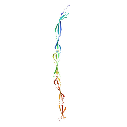
    Bacteria exploit surface proteins to adhere to other bacteria, surfaces and host cells. Such proteins need to project away from the bacterial surface and resist significant mechanical forces. SasG is a protein that forms extended fibrils on the surface of Staphylococcus aureus and promotes host adherence and biofilm formation. Here we show that although monomeric and lacking covalent cross-links, SasG maintains a highly extended conformation in solution. This extension is mediated through obligate folding cooperativity of the intrinsically disordered E domains that couple non-adjacent G5 domains thermodynamically, forming interfaces that are more stable than the domains themselves. Thus, counterintuitively, the elongation of the protein appears to be dependent on the inherent instability of its domains. The remarkable mechanical strength of SasG arises from tandemly arrayed 'clamp' motifs within the folded domains. Our findings reveal an elegant minimal solution for the assembly of monomeric mechano-resistant tethers of variable length.


  • Organizational Affiliation

    Department of Chemistry, University of Cambridge, Lensfield Road, Cambridge CB2 1EW, UK.


Macromolecules
Find similar proteins by:  (by identity cutoff)  |  3D Structure
Entity ID: 1
MoleculeChains Sequence LengthOrganismDetailsImage
Surface protein G
A, B
215Staphylococcus aureus subsp. aureus NCTC 8325Mutation(s): 0 
Gene Names: sasGSAOUHSC_02798
UniProt
Find proteins for Q2G2B2 (Staphylococcus aureus (strain NCTC 8325 / PS 47))
Explore Q2G2B2 
Go to UniProtKB:  Q2G2B2
Entity Groups  
Sequence Clusters30% Identity50% Identity70% Identity90% Identity95% Identity100% Identity
UniProt GroupQ2G2B2
Sequence Annotations
Expand
  • Reference Sequence
Small Molecules
Ligands 1 Unique
IDChains Name / Formula / InChI Key2D Diagram3D Interactions
CL
Query on CL

Download Ideal Coordinates CCD File 
C [auth A]CHLORIDE ION
Cl
VEXZGXHMUGYJMC-UHFFFAOYSA-M
Experimental Data & Validation

Experimental Data

  • Method: X-RAY DIFFRACTION
  • Resolution: 1.60 Å
  • R-Value Free: 0.249 
  • R-Value Work: 0.207 
  • R-Value Observed: 0.209 
  • Space Group: P 1
Unit Cell:
Length ( Å )Angle ( ˚ )
a = 39.861α = 84.76
b = 45.628β = 83.22
c = 60.359γ = 78.6
Software Package:
Software NamePurpose
XDSdata reduction
Aimlessdata scaling
REFMACrefinement
PDB_EXTRACTdata extraction
PHASERphasing

Structure Validation

View Full Validation Report


Entry History & Funding Information

Deposition Data


Funding OrganizationLocationGrant Number
Biotechnology and Biological Sciences Research CouncilUnited KingdomBB/J005029/1

Revision History  (Full details and data files)

  • Version 1.0: 2015-06-03
    Type: Initial release
  • Version 1.1: 2015-06-10
    Changes: Database references
  • Version 1.2: 2015-06-17
    Changes: Database references
  • Version 2.0: 2017-08-30
    Changes: Atomic model, Author supporting evidence, Derived calculations
  • Version 2.1: 2024-01-10
    Changes: Data collection, Database references, Refinement description