4WSJ | pdb_00004wsj

Crystal structure of a bacterial fucodiase in complex with 1-((1R,2R,3R,4R,5R,6R)-2,3,4-trihydroxy-5-methyl-7-azabicyclo[4.1.0]heptan-7-yl)ethan-1-one


Experimental Data Snapshot

  • Method: X-RAY DIFFRACTION
  • Resolution: 1.64 Å
  • R-Value Free: 
    0.194 (Depositor), 0.203 (DCC) 
  • R-Value Work: 
    0.164 (Depositor), 0.175 (DCC) 
  • R-Value Observed: 
    0.166 (Depositor) 

Starting Model: experimental
View more details

wwPDB Validation 3D Report Full Report

Validation slider image for 4WSJ

Ligand Structure Quality Assessment 


This is version 2.3 of the entry. See complete history

Literature

In vitroandin vivocomparative and competitive activity-based protein profiling of GH29 alpha-l-fucosidases.

Jiang, J.Kallemeijn, W.W.Wright, D.W.van den Nieuwendijk, A.M.C.H.Rohde, V.C.Folch, E.C.van den Elst, H.Florea, B.I.Scheij, S.Donker-Koopman, W.E.Verhoek, M.Li, N.Schurmann, M.Mink, D.Boot, R.G.Codee, J.D.C.van der Marel, G.A.Davies, G.J.Aerts, J.M.F.G.Overkleeft, H.S.

(2015) Chem Sci 6: 2782-false

  • DOI: https://doi.org/10.1039/c4sc03739a
  • Primary Citation Related Structures: 
    4WSJ, 4WSK

  • PubMed Abstract: 

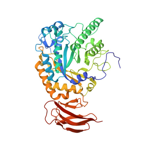
    GH29 α-l-fucosidases catalyze the hydrolysis of α-l-fucosidic linkages. Deficiency in human lysosomal α-l-fucosidase (FUCA1) leads to the recessively inherited disorder, fucosidosis. Herein we describe the development of fucopyranose-configured cyclophellitol aziridines as activity-based probes (ABPs) for selective in vitro and in vivo labeling of GH29 α-l-fucosidases from bacteria, mice and man. Crystallographic analysis on bacterial α-l-fucosidase confirms that the ABPs act by covalent modification of the active site nucleophile. Competitive activity-based protein profiling identified l-fuconojirimycin as the single GH29 α-l-fucosidase inhibitor from eight configurational isomers.


  • Organizational Affiliation
    • Leiden Institute of Chemistry , Leiden University , P. O. Box 9502 , 2300 RA Leiden , The Netherlands . Email: h.s.overkleeft@chem.leidenuniv.nl ; Email: j.m.aerts@amc.uva.nl.

Macromolecule Content 

  • Total Structure Weight: 207.39 kDa 
  • Atom Count: 15,772 
  • Modeled Residue Count: 1,761 
  • Deposited Residue Count: 1,780 
  • Unique protein chains: 1

Macromolecules

Find similar proteins by:|  3D Structure
Entity ID: 1
MoleculeChains  Sequence LengthOrganismDetailsImage
Alpha-L-fucosidase
A, B, C, D
445Bacteroides thetaiotaomicron VPI-5482Mutation(s): 0 
Gene Names: BT_2970
EC: 3.2.1.51
UniProt
Find proteins for Q8A3I4 (Bacteroides thetaiotaomicron (strain ATCC 29148 / DSM 2079 / JCM 5827 / CCUG 10774 / NCTC 10582 / VPI-5482 / E50))
Explore Q8A3I4 
Go to UniProtKB:  Q8A3I4
Entity Groups
Sequence Clusters30% Identity50% Identity70% Identity90% Identity95% Identity100% Identity
UniProt GroupQ8A3I4
Sequence Annotations
Expand
Reference Sequence

Small Molecules

Ligands 2 Unique
IDChains Name / Formula / InChI Key2D Diagram3D Interactions
3U3

Query on 3U3


Download:Ideal Coordinates CCD File
G [auth A],
J [auth B],
L [auth C],
M [auth D]
N-[(1S,2R,3R,4S,5R)-3,4,5-trihydroxy-2-methylcyclohexyl]acetamide
C9 H17 N O4
KFWZFLHIHNMMRU-ACWCSBLCSA-N
SO4

Query on SO4


Download:Ideal Coordinates CCD File
E [auth A],
F [auth A],
H [auth B],
I [auth B],
K [auth C]
SULFATE ION
O4 S
QAOWNCQODCNURD-UHFFFAOYSA-L

Experimental Data & Validation

Experimental Data

  • Method: X-RAY DIFFRACTION
  • Resolution: 1.64 Å
  • R-Value Free:  0.194 (Depositor), 0.203 (DCC) 
  • R-Value Work:  0.164 (Depositor), 0.175 (DCC) 
  • R-Value Observed: 0.166 (Depositor) 
Space Group: P 1 21 1
Unit Cell:
Length ( Å )Angle ( ˚ )
a = 55.48α = 90
b = 186.98β = 94.17
c = 98.19γ = 90
Software Package:
Software NamePurpose
Aimlessdata scaling
REFMACrefinement
PDB_EXTRACTdata extraction

Structure Validation

View Full Validation Report


Ligand Structure Quality Assessment 


Entry History 

& Funding Information

Deposition Data


Funding OrganizationLocationGrant Number
Biotechnology and Biological Sciences Research CouncilUnited KingdomA0067812

Revision History  (Full details and data files)

  • Version 1.0: 2014-11-05
    Type: Initial release
  • Version 1.1: 2015-05-27
    Changes: Database references
  • Version 2.0: 2017-08-30
    Changes: Advisory, Atomic model, Author supporting evidence, Derived calculations
  • Version 2.1: 2018-04-18
    Changes: Data collection, Database references
  • Version 2.2: 2024-01-10
    Changes: Data collection, Database references, Refinement description
  • Version 2.3: 2024-10-23
    Changes: Structure summary