4WRQ | pdb_00004wrq

Crystal Structure of 14-3-3 zeta with Chibby peptide


Experimental Data Snapshot

  • Method: X-RAY DIFFRACTION
  • Resolution: 2.41 Å
  • R-Value Free: 
    0.256 (Depositor), 0.259 (DCC) 
  • R-Value Work: 
    0.203 (Depositor), 0.206 (DCC) 
  • R-Value Observed: 
    0.207 (Depositor) 

Starting Model: experimental
View more details

wwPDB Validation 3D Report Full Report

Validation slider image for 4WRQ

This is version 1.4 of the entry. See complete history

Literature

Structural Analysis of the 14-3-3 zeta /Chibby Interaction Involved in Wnt/ beta-Catenin Signaling.

Killoran, R.C.Fan, J.Yang, D.Shilton, B.H.Choy, W.Y.

(2015) PLoS One 10: e0123934-e0123934

  • DOI: https://doi.org/10.1371/journal.pone.0123934
  • Primary Citation Related Structures: 
    4WRQ

  • PubMed Abstract: 

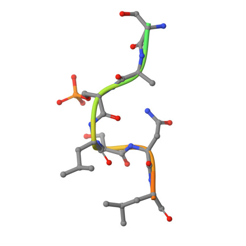
    The partially disordered Chibby (Cby) is a conserved nuclear protein that antagonizes the Wnt/β-catenin signaling pathway. By competing with the Tcf/Lef family proteins for binding to β-catenin, Cby abrogates the β-catenin-mediated transcription of Wnt signaling genes. Additionally, upon phosphorylation on S20 by the kinase Akt, Cby forms a complex with 14-3-3 to facilitate the nuclear export of β-catenin, which represents another crucial mechanism for the regulation of Wnt signaling. To obtain a mechanistic understanding of the 14-3-3/Cby interaction, we have extensively characterized the complex using X-ray crystallography, nuclear magnetic resonance (NMR) spectroscopy, and isothermal titration calorimetry (ITC). The crystal structure of the human 14-3-3ζ/Cby protein-peptide complex reveals a canonical binding mode; however the residue at the +2 position from the phosphorylated serine is shown to be uniquely oriented relative to other solved structures of 14-3-3 complexes. Our ITC results illustrate that although the phosphorylation of S20 is essential for Cby to recognize 14-3-3, residues flanking the phosphorylation site also contribute to the binding affinity. However, as is commonly observed in other 14-3-3/phosphopeptide crystal structures, residues of Cby flanking the 14-3-3 binding motif lack observable electron density. To obtain a more detailed binding interface, we have completed the backbone NMR resonance assignment of 14-3-3ζ. NMR titration experiments reveal that residues outside of the 14-3-3 conserved binding cleft, namely a flexible loop consisting of residues 203-210, are also involved in binding Cby. By using a combined X-ray and NMR approach, we have dissected the molecular basis of the 14-3-3/Cby interaction.


  • Organizational Affiliation
    • Department of Biochemistry, The University of Western Ontario, London, Ontario N6A 5C1, Canada.

Macromolecule Content 

  • Total Structure Weight: 59.85 kDa 
  • Atom Count: 3,857 
  • Modeled Residue Count: 464 
  • Deposited Residue Count: 528 
  • Unique protein chains: 2

Macromolecules

Find similar proteins by:|  3D Structure
Entity ID: 1
MoleculeChains  Sequence LengthOrganismDetailsImage
14-3-3 protein zeta/delta
A, B
246Homo sapiensMutation(s): 0 
Gene Names: YWHAZ
UniProt & NIH Common Fund Data Resources
Find proteins for P63104 (Homo sapiens)
Explore P63104 
Go to UniProtKB:  P63104
PHAROS:  P63104
GTEx:  ENSG00000164924 
Entity Groups
Sequence Clusters30% Identity50% Identity70% Identity90% Identity95% Identity100% Identity
UniProt GroupP63104
Sequence Annotations
Expand
Reference Sequence
Find similar proteins by:  Sequence   |   3D Structure  
Entity ID: 2
MoleculeChains  Sequence LengthOrganismDetailsImage
Chibby
C, D
18Homo sapiensMutation(s): 0 
UniProt & NIH Common Fund Data Resources
Find proteins for Q9Y3M2 (Homo sapiens)
Explore Q9Y3M2 
Go to UniProtKB:  Q9Y3M2
PHAROS:  Q9Y3M2
GTEx:  ENSG00000100211 
Entity Groups
Sequence Clusters30% Identity50% Identity70% Identity90% Identity95% Identity100% Identity
UniProt GroupQ9Y3M2
Sequence Annotations
Expand
Reference Sequence

Small Molecules

Modified Residues  1 Unique
IDChains TypeFormula2D DiagramParent
SEP
Query on SEP
C, D
L-PEPTIDE LINKINGC3 H8 N O6 PSER

Experimental Data & Validation

Experimental Data

  • Method: X-RAY DIFFRACTION
  • Resolution: 2.41 Å
  • R-Value Free:  0.256 (Depositor), 0.259 (DCC) 
  • R-Value Work:  0.203 (Depositor), 0.206 (DCC) 
  • R-Value Observed: 0.207 (Depositor) 
Space Group: P 21 21 21
Unit Cell:
Length ( Å )Angle ( ˚ )
a = 70.83α = 90
b = 71.96β = 90
c = 130.99γ = 90
Software Package:
Software NamePurpose
PHENIXrefinement
PDB_EXTRACTdata extraction

Structure Validation

View Full Validation Report


Entry History 

& Funding Information

Deposition Data


Funding OrganizationLocationGrant Number
Canadian Institutes of Health Research (CIHR)CanadaMOP 74679

Revision History  (Full details and data files)

  • Version 1.0: 2015-05-06
    Type: Initial release
  • Version 1.1: 2017-09-13
    Changes: Author supporting evidence, Derived calculations, Other, Source and taxonomy, Structure summary
  • Version 1.2: 2020-01-08
    Changes: Author supporting evidence
  • Version 1.3: 2023-09-27
    Changes: Data collection, Database references, Refinement description
  • Version 1.4: 2024-10-30
    Changes: Structure summary