4WOY | pdb_00004woy

Crystal structure and functional analysis of MiD49, a receptor for the mitochondrial fission protein Drp1


Experimental Data Snapshot

  • Method: X-RAY DIFFRACTION
  • Resolution: 2.40 Å
  • R-Value Free: 
    0.243 (Depositor), 0.246 (DCC) 
  • R-Value Work: 
    0.206 (Depositor), 0.212 (DCC) 
  • R-Value Observed: 
    0.207 (Depositor) 

wwPDB Validation 3D Report Full Report

Validation slider image for 4WOY

This is version 1.2 of the entry. See complete history

Literature

Crystal structure and functional analysis of MiD49, a receptor for the mitochondrial fission protein Drp1.

Loson, O.C.Meng, S.Ngo, H.Liu, R.Kaiser, J.T.Chan, D.C.

(2015) Protein Sci 24: 386-394

  • DOI: https://doi.org/10.1002/pro.2629
  • Primary Citation Related Structures: 
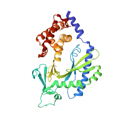
    4WOY

  • PubMed Abstract: 

    Mitochondrial fission requires recruitment of dynamin-related protein 1 (Drp1) to the mitochondrial surface, where assembly leads to activation of its GTP-dependent scission function. MiD49 and MiD51 are two receptors on the mitochondrial outer membrane that can recruit Drp1 to facilitate mitochondrial fission. Structural studies indicated that MiD51 has a variant nucleotidyl transferase fold that binds an ADP co-factor essential for activation of Drp1 function. MiD49 shares sequence homology with MiD51 and regulates Drp1 function. However, it is unknown if MiD49 binds an analogous co-factor. Because MiD49 does not readily crystallize, we used structural predictions and biochemical screening to identify a surface entropy reduction mutant that facilitated crystallization. Using molecular replacement, we determined the atomic structure of MiD49 to 2.4 Å. Like MiD51, MiD49 contains a nucleotidyl transferase domain; however, the electron density provides no evidence for a small-molecule ligand. Structural changes in the putative nucleotide-binding pocket make MiD49 incompatible with an extended ligand like ADP, and critical nucleotide-binding residues found in MiD51 are not conserved. MiD49 contains a surface loop that physically interacts with Drp1 and is necessary for Drp1 recruitment to the mitochondrial surface. Our results suggest a structural basis for the differential regulation of MiD51- versus MiD49-mediated fission.


  • Organizational Affiliation
    • Division of Biology and Biological Engineering, California Institute of Technology, Pasadena, California, 91125.

Macromolecule Content 

  • Total Structure Weight: 73.16 kDa 
  • Atom Count: 5,337 
  • Modeled Residue Count: 658 
  • Deposited Residue Count: 658 
  • Unique protein chains: 1

Macromolecules

Find similar proteins by:|  3D Structure
Entity ID: 1
MoleculeChains  Sequence LengthOrganismDetailsImage
Mitochondrial dynamics protein MID49
A, B
329Mus musculusMutation(s): 1 
Gene Names: Mief2Gm11Mid49Smcr7
UniProt
Find proteins for Q5NCS9 (Mus musculus)
Explore Q5NCS9 
Go to UniProtKB:  Q5NCS9
Entity Groups
Sequence Clusters30% Identity50% Identity70% Identity90% Identity95% Identity100% Identity
UniProt GroupQ5NCS9
Sequence Annotations
Expand
Reference Sequence

Experimental Data & Validation

Experimental Data

  • Method: X-RAY DIFFRACTION
  • Resolution: 2.40 Å
  • R-Value Free:  0.243 (Depositor), 0.246 (DCC) 
  • R-Value Work:  0.206 (Depositor), 0.212 (DCC) 
  • R-Value Observed: 0.207 (Depositor) 
Space Group: C 2 2 21
Unit Cell:
Length ( Å )Angle ( ˚ )
a = 100.674α = 90
b = 153.39β = 90
c = 109.015γ = 90
Software Package:
Software NamePurpose
PHENIXrefinement

Structure Validation

View Full Validation Report


Entry History 

Deposition Data

Revision History  (Full details and data files)

  • Version 1.0: 2015-01-28
    Type: Initial release
  • Version 1.1: 2015-03-04
    Changes: Database references
  • Version 1.2: 2023-12-27
    Changes: Data collection, Database references, Derived calculations, Other, Refinement description, Source and taxonomy, Structure summary