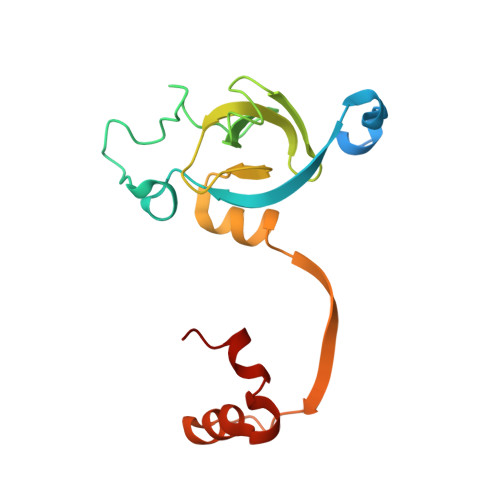
4WLK

Stationary Phase Survival Protein YuiC from B.subtilis complexed with reaction product


Experimental Data Snapshot

  • Method: X-RAY DIFFRACTION
  • Resolution: 2.03 Å
  • R-Value Free: 0.205 
  • R-Value Work: 0.172 
  • R-Value Observed: 0.173 

Starting Model: experimental
View more details

wwPDB Validation   3D Report Full Report


Ligand Structure Quality Assessment 


This is version 1.2 of the entry. See complete history


Literature

Structure of the stationary phase survival protein YuiC from B.subtilis.

Quay, D.H.Cole, A.R.Cryar, A.Thalassinos, K.Williams, M.A.Bhakta, S.Keep, N.H.

(2015) BMC Struct Biol 15: 12-12

  • DOI: https://doi.org/10.1186/s12900-015-0039-z
  • Primary Citation of Related Structures:  
    4WJT, 4WLI, 4WLK

  • PubMed Abstract: 

    Stationary phase survival proteins (Sps) were found in Firmicutes as having analogous domain compositions, and in some cases genome context, as the resuscitation promoting factors of Actinobacteria, but with a different putative peptidoglycan cleaving domain. The first structure of a Firmicute Sps protein YuiC from B. subtilis, is found to be a stripped down version of the cell-wall peptidoglycan hydrolase MltA. The YuiC structures are of a domain swapped dimer, although some monomer is also found in solution. The protein crystallised in the presence of pentasaccharide shows a 1,6-anhydrodisaccharide sugar product, indicating that YuiC cleaves the sugar backbone to form an anhydro product at least on lengthy incubation during crystallisation. The structural simplification of MltA in Sps proteins is analogous to that of the resuscitation promoting factor domains of Actinobacteria, which are stripped down versions of lysozyme and soluble lytic transglycosylase proteins.


  • Organizational Affiliation

    Institute for Structural and Molecular Biology, Crystallography, Department of Biological Sciences, Birkbeck University of London, Malet Street, London, WC1E 7HX, UK. d.quay@mail.cryst.bbk.ac.uk.


Macromolecules
Find similar proteins by:  (by identity cutoff)  |  3D Structure
Entity ID: 1
MoleculeChains Sequence LengthOrganismDetailsImage
YuiC
A, B
166Bacillus subtilis subsp. subtilis str. 168Mutation(s): 0 
Gene Names: yuiCBSU32070
UniProt
Find proteins for O32108 (Bacillus subtilis (strain 168))
Explore O32108 
Go to UniProtKB:  O32108
Entity Groups  
Sequence Clusters30% Identity50% Identity70% Identity90% Identity95% Identity100% Identity
UniProt GroupO32108
Sequence Annotations
Expand
  • Reference Sequence
Small Molecules
Ligands 1 Unique
IDChains Name / Formula / InChI Key2D Diagram3D Interactions
3QL
Query on 3QL

Download Ideal Coordinates CCD File 
C [auth A],
D [auth B]
N-[(1R,2S,3R,4R,5R)-2-[(2S,3R,4R,5S,6R)-3-acetamido-6-(hydroxymethyl)-4,5-bis(oxidanyl)oxan-2-yl]oxy-3-oxidanyl-6,8-dioxabicyclo[3.2.1]octan-4-yl]ethanamide
C16 H26 N2 O10
UVQRTGABPMLDOA-KSKNGZLJSA-N
Experimental Data & Validation

Experimental Data

  • Method: X-RAY DIFFRACTION
  • Resolution: 2.03 Å
  • R-Value Free: 0.205 
  • R-Value Work: 0.172 
  • R-Value Observed: 0.173 
  • Space Group: H 3
Unit Cell:
Length ( Å )Angle ( ˚ )
a = 147.21α = 90
b = 147.21β = 90
c = 37.87γ = 120
Software Package:
Software NamePurpose
PHENIXrefinement
PHASERphasing
Aimlessdata scaling
XDSdata reduction

Structure Validation

View Full Validation Report


Ligand Structure Quality Assessment 


Entry History 

Deposition Data

Revision History  (Full details and data files)

  • Version 1.0: 2015-07-08
    Type: Initial release
  • Version 1.1: 2016-09-21
    Changes: Database references
  • Version 1.2: 2024-01-10
    Changes: Data collection, Database references, Refinement description