4WF0 | pdb_00004wf0

Crystal Structure of iLID - an Improved Light-Inducible Dimer


Experimental Data Snapshot

  • Method: X-RAY DIFFRACTION
  • Resolution: 1.95 Å
  • R-Value Free: 
    0.244 (Depositor), 0.255 (DCC) 
  • R-Value Work: 
    0.231 (Depositor), 0.246 (DCC) 
  • R-Value Observed: 
    0.232 (Depositor) 

Starting Model: experimental
View more details

wwPDB Validation 3D Report Full Report

Validation slider image for 4WF0

Ligand Structure Quality Assessment 


This is version 1.4 of the entry. See complete history

Literature

Engineering an improved light-induced dimer (iLID) for controlling the localization and activity of signaling proteins.

Guntas, G.Hallett, R.A.Zimmerman, S.P.Williams, T.Yumerefendi, H.Bear, J.E.Kuhlman, B.

(2015) Proc Natl Acad Sci U S A 112: 112-117

  • DOI: https://doi.org/10.1073/pnas.1417910112
  • Primary Citation Related Structures: 
    4WF0

  • PubMed Abstract: 

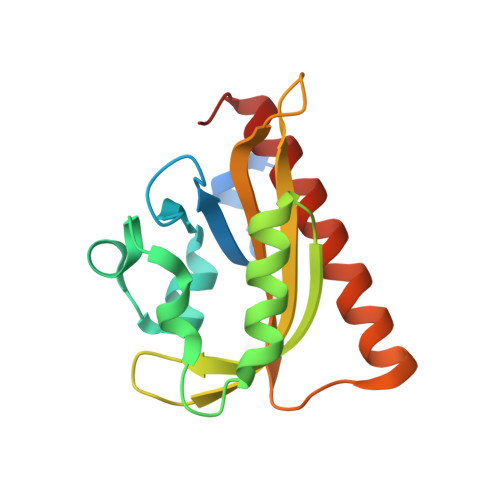
    The discovery of light-inducible protein-protein interactions has allowed for the spatial and temporal control of a variety of biological processes. To be effective, a photodimerizer should have several characteristics: it should show a large change in binding affinity upon light stimulation, it should not cross-react with other molecules in the cell, and it should be easily used in a variety of organisms to recruit proteins of interest to each other. To create a switch that meets these criteria we have embedded the bacterial SsrA peptide in the C-terminal helix of a naturally occurring photoswitch, the light-oxygen-voltage 2 (LOV2) domain from Avena sativa. In the dark the SsrA peptide is sterically blocked from binding its natural binding partner, SspB. When activated with blue light, the C-terminal helix of the LOV2 domain undocks from the protein, allowing the SsrA peptide to bind SspB. Without optimization, the switch exhibited a twofold change in binding affinity for SspB with light stimulation. Here, we describe the use of computational protein design, phage display, and high-throughput binding assays to create an improved light inducible dimer (iLID) that changes its affinity for SspB by over 50-fold with light stimulation. A crystal structure of iLID shows a critical interaction between the surface of the LOV2 domain and a phenylalanine engineered to more tightly pin the SsrA peptide against the LOV2 domain in the dark. We demonstrate the functional utility of the switch through light-mediated subcellular localization in mammalian cell culture and reversible control of small GTPase signaling.


  • Organizational Affiliation
    • Department of Biochemistry & Biophysics.

Macromolecule Content 

  • Total Structure Weight: 37.52 kDa 
  • Atom Count: 2,711 
  • Modeled Residue Count: 288 
  • Deposited Residue Count: 316 
  • Unique protein chains: 1

Macromolecules

Find similar proteins by:|  3D Structure
Entity ID: 1
MoleculeChains  Sequence LengthOrganismDetailsImage
NPH1-1
A, B
158Avena sativaMutation(s): 11 
Gene Names: NPH1-1
EC: 2.7.11.1
UniProt
Find proteins for O49003 (Avena sativa)
Explore O49003 
Go to UniProtKB:  O49003
Entity Groups
Sequence Clusters30% Identity50% Identity70% Identity90% Identity95% Identity100% Identity
UniProt GroupO49003
Sequence Annotations
Expand
Reference Sequence

Experimental Data & Validation

Experimental Data

  • Method: X-RAY DIFFRACTION
  • Resolution: 1.95 Å
  • R-Value Free:  0.244 (Depositor), 0.255 (DCC) 
  • R-Value Work:  0.231 (Depositor), 0.246 (DCC) 
  • R-Value Observed: 0.232 (Depositor) 
Space Group: P 21 21 21
Unit Cell:
Length ( Å )Angle ( ˚ )
a = 62.156α = 90
b = 70.339β = 90
c = 80.378γ = 90
Software Package:
Software NamePurpose
PHENIXrefinement
SCALAdata scaling
XDSdata reduction

Structure Validation

View Full Validation Report


Ligand Structure Quality Assessment 


Entry History 

& Funding Information

Deposition Data


Funding OrganizationLocationGrant Number
National Institutes of Health/National Institute of General Medical Sciences (NIH/NIGMS)United StatesGM093208

Revision History  (Full details and data files)

  • Version 1.0: 2014-12-24
    Type: Initial release
  • Version 1.1: 2015-01-07
    Changes: Database references
  • Version 1.2: 2015-01-14
    Changes: Database references
  • Version 1.3: 2019-12-04
    Changes: Author supporting evidence, Database references, Derived calculations, Source and taxonomy
  • Version 1.4: 2023-09-27
    Changes: Data collection, Database references, Refinement description