4WEL | pdb_00004wel

Crystal structure of Pseudomonas aeruginosa PBP3 with SMC-3176


Experimental Data Snapshot

  • Method: X-RAY DIFFRACTION
  • Resolution: 1.99 Å
  • R-Value Free: 
    0.220 (Depositor), 0.219 (DCC) 
  • R-Value Work: 
    0.190 (Depositor) 
  • R-Value Observed: 
    0.191 (Depositor) 

Starting Model: experimental
View more details

wwPDB Validation 3D Report Full Report

Validation slider image for 4WEL

Ligand Structure Quality Assessment 


This is version 1.3 of the entry. See complete history

Literature

SAR and Structural Analysis of Siderophore-Conjugated Monocarbam Inhibitors of Pseudomonas aeruginosa PBP3.

Murphy-Benenato, K.E.Dangel, B.Davis, H.E.Durand-Reville, T.F.Ferguson, A.D.Gao, N.Jahic, H.Mueller, J.P.Manyak, E.L.Quiroga, O.Rooney, M.Sha, L.Sylvester, M.Wu, F.Zambrowski, M.Zhao, S.X.

(2015) ACS Med Chem Lett 6: 537-542

  • DOI: https://doi.org/10.1021/acsmedchemlett.5b00026
  • Primary Citation Related Structures: 
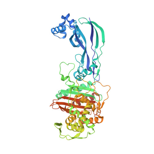
    4WEJ, 4WEK, 4WEL

  • PubMed Abstract: 

    A main challenge in the development of new agents for the treatment of Pseudomonas aeruginosa infections is the identification of chemotypes that efficiently penetrate the cell envelope and are not susceptible to established resistance mechanisms. Siderophore-conjugated monocarbams are attractive because of their ability to hijack the bacteria's iron uptake machinery for transport into the periplasm and their inherent stability to metallo-β-lactamases. Through development of the SAR we identified a number of modifications to the scaffold that afforded active anti-P. aeruginosa agents with good physicochemical properties. Through crystallographic efforts we gained a better understanding into how these compounds bind to the target penicillin binding protein PBP3 and factors to consider for future design.


  • Organizational Affiliation
    • Infection Innovative Medicines, AstraZeneca R&D Boston , 35 Gatehouse Drive, Waltham, Massachusetts 02451, United States.

Macromolecule Content 

  • Total Structure Weight: 59.11 kDa 
  • Atom Count: 4,087 
  • Modeled Residue Count: 500 
  • Deposited Residue Count: 538 
  • Unique protein chains: 1

Macromolecules

Find similar proteins by:|  3D Structure
Entity ID: 1
MoleculeChains  Sequence LengthOrganismDetailsImage
Penicillin-binding protein 3538Pseudomonas aeruginosa PA14Mutation(s): 0 
Gene Names: CIA_00399
UniProt
Find proteins for A0A0M3KKZ3 (Pseudomonas aeruginosa PA14)
Explore A0A0M3KKZ3 
Go to UniProtKB:  A0A0M3KKZ3
Entity Groups
Sequence Clusters30% Identity50% Identity70% Identity90% Identity95% Identity100% Identity
UniProt GroupA0A0M3KKZ3
Sequence Annotations
Expand
Reference Sequence

Small Molecules

Ligands 1 Unique
IDChains Name / Formula / InChI Key2D Diagram3D Interactions
3LE

Query on 3LE


Download:Ideal Coordinates CCD File
B [auth A](3S,4S,7Z)-7-(2-amino-1,3-thiazol-4-yl)-4-formyl-1-[({3-(5-hydroxy-4-oxo-3,4-dihydropyridin-2-yl)-4-[3-(methylsulfonyl)propyl]-5-oxo-4,5-dihydro-1H-1,2,4-triazol-1-yl}sulfonyl)amino]-3,10,10-trimethyl-1,6-dioxo-9-oxa-2,5,8-triazaundec-7-en-11-oic acid
C25 H32 N10 O13 S3
KOZNTWBICKFGGR-BECCELLYSA-N

Experimental Data & Validation

Experimental Data

  • Method: X-RAY DIFFRACTION
  • Resolution: 1.99 Å
  • R-Value Free:  0.220 (Depositor), 0.219 (DCC) 
  • R-Value Work:  0.190 (Depositor) 
  • R-Value Observed: 0.191 (Depositor) 
Space Group: P 21 21 21
Unit Cell:
Length ( Å )Angle ( ˚ )
a = 68.442α = 90
b = 83.337β = 90
c = 89.411γ = 90
Software Package:
Software NamePurpose
BUSTERrefinement
Cootmodel building
MOLREPphasing
XDSdata reduction
SCALAdata scaling

Structure Validation

View Full Validation Report


Ligand Structure Quality Assessment 


Entry History 

Deposition Data

Revision History  (Full details and data files)

  • Version 1.0: 2015-04-22
    Type: Initial release
  • Version 1.1: 2015-06-10
    Changes: Database references
  • Version 1.2: 2023-09-27
    Changes: Data collection, Database references, Derived calculations, Other, Refinement description, Source and taxonomy, Structure summary
  • Version 1.3: 2024-11-13
    Changes: Structure summary