4WE2 | pdb_00004we2

Donor strand complemented FaeG of F4ab fimbriae


Experimental Data Snapshot

  • Method: X-RAY DIFFRACTION
  • Resolution: 1.50 Å
  • R-Value Free: 
    0.214 (Depositor), 0.235 (DCC) 
  • R-Value Work: 
    0.175 (Depositor) 
  • R-Value Observed: 
    0.177 (Depositor) 

Starting Model: experimental
View more details

wwPDB Validation 3D Report Full Report

Validation slider image for 4WE2

This is version 1.3 of the entry. See complete history

Literature

Structural and Functional Insight into the Carbohydrate Receptor Binding of F4 Fimbriae-producing Enterotoxigenic Escherichia coli.

Moonens, K.Van den Broeck, I.De Kerpel, M.Deboeck, F.Raymaekers, H.Remaut, H.De Greve, H.

(2015) J Biological Chem 290: 8409-8419

  • DOI: https://doi.org/10.1074/jbc.M114.618595
  • Primary Citation Related Structures: 
    4WE2, 4WEI

  • PubMed Abstract: 

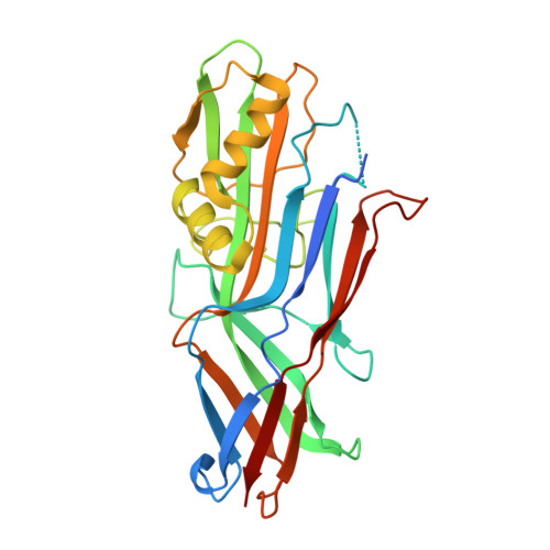
    Enterotoxigenic Escherichia coli (ETEC) strains are important causes of intestinal disease in humans and lead to severe production losses in animal farming. A range of fimbrial adhesins in ETEC strains determines host and tissue tropism. ETEC strains expressing F4 fimbriae are associated with neonatal and post-weaning diarrhea in piglets. Three naturally occurring variants of F4 fimbriae (F4ab, F4ac, and F4ad) exist that differ in the primary sequence of their major adhesive subunit FaeG, and each features a related yet distinct receptor binding profile. Here the x-ray structure of FaeGad bound to lactose provides the first structural insight into the receptor specificity and mode of binding by the poly-adhesive F4 fimbriae. A small D'-D″-α1-α2 subdomain grafted on the immunoglobulin-like core of FaeG hosts the carbohydrate binding site. Two short amino acid stretches Phe(150)-Glu(152) and Val(166)-Glu(170) of FaeGad bind the terminal galactose in the lactosyl unit and provide affinity and specificity to the interaction. A hemagglutination-based assay with E. coli expressing mutant F4ad fimbriae confirmed the elucidated co-complex structure. Interestingly, the crucial D'-α1 loop that borders the FaeGad binding site adopts a different conformation in the two other FaeG variants and hints at a heterogeneous binding pocket among the FaeG serotypes.


  • Organizational Affiliation
    • From the Structural and Molecular Microbiology, VIB Structural Biology Research Center, 1050 Brussels, the Structural Biology Brussels, Vrije Universiteit Brussel, Pleinlaan 2, 1050 Brussels, and.

Macromolecule Content 

  • Total Structure Weight: 29.13 kDa 
  • Atom Count: 2,238 
  • Modeled Residue Count: 260 
  • Deposited Residue Count: 277 
  • Unique protein chains: 1

Macromolecules

Find similar proteins by:|  3D Structure
Entity ID: 1
MoleculeChains  Sequence LengthOrganismDetailsImage
K88 fimbrial protein AB277Escherichia coliMutation(s): 0 
Gene Names: faeG
UniProt
Find proteins for P02970 (Escherichia coli)
Explore P02970 
Go to UniProtKB:  P02970
Entity Groups
Sequence Clusters30% Identity50% Identity70% Identity90% Identity95% Identity100% Identity
UniProt GroupP02970
Sequence Annotations
Expand
Reference Sequence

Experimental Data & Validation

Experimental Data

  • Method: X-RAY DIFFRACTION
  • Resolution: 1.50 Å
  • R-Value Free:  0.214 (Depositor), 0.235 (DCC) 
  • R-Value Work:  0.175 (Depositor) 
  • R-Value Observed: 0.177 (Depositor) 
Space Group: P 1 21 1
Unit Cell:
Length ( Å )Angle ( ˚ )
a = 53.639α = 90
b = 49.026β = 109.21
c = 54.468γ = 90
Software Package:
Software NamePurpose
REFMACrefinement
XDSdata reduction
SCALAdata scaling
PHASERphasing

Structure Validation

View Full Validation Report


Entry History 

& Funding Information

Deposition Data


Funding OrganizationLocationGrant Number
FWOBelgiumG030411N
FWO - HerculesBelgiumUABR/09/005

Revision History  (Full details and data files)

  • Version 1.0: 2015-02-04
    Type: Initial release
  • Version 1.1: 2015-02-11
    Changes: Database references
  • Version 1.2: 2015-04-08
    Changes: Database references
  • Version 1.3: 2024-01-10
    Changes: Data collection, Database references, Refinement description