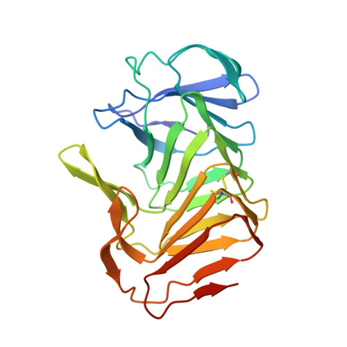
4W8S | pdb_00004w8s

Crystal structure of truncated hemolysin A Q125S/Y134S from P. mirabilis at 1.5 Angstroms resolution


Experimental Data Snapshot

  • Method: X-RAY DIFFRACTION
  • Resolution: 1.51 Å
  • R-Value Free: 
    0.201 (Depositor), 0.202 (DCC) 
  • R-Value Work: 
    0.176 (Depositor), 0.179 (DCC) 
  • R-Value Observed: 
    0.177 (Depositor) 

Starting Model: experimental
View more details

wwPDB Validation 3D Report Full Report

Validation slider image for 4W8S

This is version 1.5 of the entry. See complete history

Literature

Crystal structure of truncated hemolysin A Q125S/Y134S from P. mirabilis at 1.5 Angstroms resolution

Novak, W.R.P.Glasgow, E.Thompson, J.R.Weaver, T.M.

To be published.

Macromolecule Content 

  • Total Structure Weight: 25.36 kDa 
  • Atom Count: 2,059 
  • Modeled Residue Count: 234 
  • Deposited Residue Count: 242 
  • Unique protein chains: 1

Macromolecules

Find similar proteins by:|  3D Structure
Entity ID: 1
MoleculeChains  Sequence LengthOrganismDetailsImage
Hemolysin242Proteus mirabilisMutation(s): 2 
Gene Names: hpmA
UniProt
Find proteins for P16466 (Proteus mirabilis)
Explore P16466 
Go to UniProtKB:  P16466
Entity Groups
Sequence Clusters30% Identity50% Identity70% Identity90% Identity95% Identity100% Identity
UniProt GroupP16466
Sequence Annotations
Expand
Reference Sequence

Experimental Data & Validation

Experimental Data

  • Method: X-RAY DIFFRACTION
  • Resolution: 1.51 Å
  • R-Value Free:  0.201 (Depositor), 0.202 (DCC) 
  • R-Value Work:  0.176 (Depositor), 0.179 (DCC) 
  • R-Value Observed: 0.177 (Depositor) 
Space Group: P 21 21 2
Unit Cell:
Length ( Å )Angle ( ˚ )
a = 56.338α = 90
b = 120.153β = 90
c = 34.01γ = 90
Software Package:
Software NamePurpose
PHENIXrefinement

Structure Validation

View Full Validation Report


Entry History 

& Funding Information

Deposition Data


Funding OrganizationLocationGrant Number
National Science Foundation (NSF, United States)United StatesMCB1050435

Revision History  (Full details and data files)

  • Version 1.0: 2015-10-07
    Type: Initial release
  • Version 1.1: 2017-06-21
    Changes: Structure summary
  • Version 1.2: 2017-09-20
    Changes: Author supporting evidence
  • Version 1.3: 2019-11-27
    Changes: Author supporting evidence
  • Version 1.4: 2023-09-27
    Changes: Data collection, Database references, Refinement description
  • Version 1.5: 2024-11-06
    Changes: Structure summary