4W7R | pdb_00004w7r

Crystal Structure of Full-Length Split GFP Mutant E124H/K126H Copper Mediated Dimer, P 21 Space Group


Experimental Data Snapshot

  • Method: X-RAY DIFFRACTION
  • Resolution: 1.80 Å
  • R-Value Free: 
    0.253 (Depositor), 0.253 (DCC) 
  • R-Value Work: 
    0.223 (Depositor), 0.224 (DCC) 
  • R-Value Observed: 
    0.226 (Depositor) 

Starting Model: experimental
View more details

wwPDB Validation 3D Report Full Report

Validation slider image for 4W7R

This is version 1.6 of the entry. See complete history

Literature

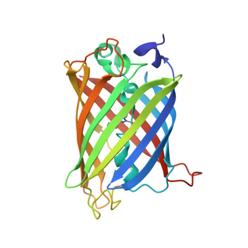
A Suite of Engineered GFP Molecules for Oligomeric Scaffolding.

Leibly, D.J.Arbing, M.A.Pashkov, I.DeVore, N.Waldo, G.S.Terwilliger, T.C.Yeates, T.O.

(2015) Structure 23: 1754-1768

  • DOI: https://doi.org/10.1016/j.str.2015.07.008
  • Primary Citation Related Structures: 
    4W69, 4W6A, 4W6B, 4W6C, 4W6D, 4W6F, 4W6G, 4W6H, 4W6I, 4W6J, 4W6K, 4W6L, 4W6M, 4W6N, 4W6O, 4W6P, 4W6R, 4W6S, 4W6T, 4W6U, 4W72, 4W73, 4W74, 4W75, 4W76, 4W77, 4W7A, 4W7C, 4W7D, 4W7E, 4W7F, 4W7R, 4W7X

  • PubMed Abstract: 

    Applications ranging from synthetic biology to protein crystallization could be advanced by facile systems for connecting multiple proteins together in predefined spatial relationships. One approach to this goal is to engineer many distinct assembly forms of a single carrier protein or scaffold, to which other proteins of interest can then be readily attached. In this work we chose GFP as a scaffold and engineered many alternative oligomeric forms, driven by either specific disulfide bond formation or metal ion addition. We generated a wide range of spatial arrangements of GFP subunits from 11 different oligomeric variants, and determined their X-ray structures in a total of 33 distinct crystal forms. Some of the oligomeric GFP variants show geometric polymorphism depending on conditions, while others show considerable geometric rigidity. Potential future applications of this system are discussed.


  • Organizational Affiliation
    • Department of Chemistry and Biochemistry, University of California, Los Angeles, CA 90095, USA; UCLA-DOE Institute of Genomics and Proteomics, University of California, Los Angeles, CA 90095, USA.

Macromolecule Content 

  • Total Structure Weight: 105.29 kDa 
  • Atom Count: 7,625 
  • Modeled Residue Count: 903 
  • Deposited Residue Count: 916 
  • Unique protein chains: 1

Macromolecules

Find similar proteins by:|  3D Structure
Entity ID: 1
MoleculeChains  Sequence LengthOrganismDetailsImage
fluorescent protein E124H/K126C
A, B, C, D
229synthetic constructMutation(s): 0 

Small Molecules

Ligands 2 Unique
IDChains Name / Formula / InChI Key2D Diagram3D Interactions
CU

Query on CU


Download:Ideal Coordinates CCD File
E [auth A],
M [auth C]
COPPER (II) ION
Cu
JPVYNHNXODAKFH-UHFFFAOYSA-N
EDO

Query on EDO


Download:Ideal Coordinates CCD File
F [auth A]
G [auth A]
H [auth A]
I [auth B]
J [auth B]
F [auth A],
G [auth A],
H [auth A],
I [auth B],
J [auth B],
K [auth B],
L [auth B],
N [auth C],
O [auth C],
P [auth D],
Q [auth D],
R [auth D],
S [auth D],
T [auth D]
1,2-ETHANEDIOL
C2 H6 O2
LYCAIKOWRPUZTN-UHFFFAOYSA-N
Modified Residues  1 Unique
IDChains TypeFormula2D DiagramParent
CRO
Query on CRO
A, B, C, D
L-PEPTIDE LINKINGC15 H17 N3 O5THR, TYR, GLY

Experimental Data & Validation

Experimental Data

  • Method: X-RAY DIFFRACTION
  • Resolution: 1.80 Å
  • R-Value Free:  0.253 (Depositor), 0.253 (DCC) 
  • R-Value Work:  0.223 (Depositor), 0.224 (DCC) 
  • R-Value Observed: 0.226 (Depositor) 
Space Group: P 1 21 1
Unit Cell:
Length ( Å )Angle ( ˚ )
a = 62.67α = 90
b = 87.19β = 90.01
c = 92.07γ = 90
Software Package:
Software NamePurpose
PHENIXrefinement
XDSdata reduction
XSCALEdata scaling
PHASERphasing

Structure Validation

View Full Validation Report


Entry History 

& Funding Information

Deposition Data


Funding OrganizationLocationGrant Number
National Institutes of Health/National Institute of General Medical Sciences (NIH/NIGMS)United StatesGM098177

Revision History  (Full details and data files)

  • Version 1.0: 2015-03-04
    Type: Initial release
  • Version 1.1: 2016-01-27
    Changes: Database references
  • Version 1.2: 2017-09-27
    Changes: Author supporting evidence, Database references, Derived calculations
  • Version 1.3: 2019-12-25
    Changes: Author supporting evidence
  • Version 1.4: 2023-09-27
    Changes: Data collection, Database references, Derived calculations, Refinement description
  • Version 1.5: 2023-11-15
    Changes: Data collection
  • Version 1.6: 2024-10-30
    Changes: Structure summary