4W5K | pdb_00004w5k

Structure of a mitochondrial aspartate aminotransferase from Trypanosoma brucei, K237A mutant


Experimental Data Snapshot

  • Method: X-RAY DIFFRACTION
  • Resolution: 1.70 Å
  • R-Value Free: 
    0.183 (Depositor), 0.191 (DCC) 
  • R-Value Work: 
    0.153 (Depositor) 
  • R-Value Observed: 
    0.154 (Depositor) 

Starting Model: experimental
View more details

wwPDB Validation 3D Report Full Report

Validation slider image for 4W5K

Ligand Structure Quality Assessment 


This is version 2.0 of the entry. See complete history

Literature

Structures of aspartate aminotransferases from Trypanosoma brucei, Leishmania major and Giardia lamblia.

Abendroth, J.Choi, R.Wall, A.Clifton, M.C.Lukacs, C.M.Staker, B.L.Van Voorhis, W.Myler, P.Lorimer, D.D.Edwards, T.E.

(2015) Acta Crystallogr F Struct Biol Commun 71: 566-571

  • DOI: https://doi.org/10.1107/S2053230X15001831
  • Primary Citation Related Structures: 
    3MEB, 4EU1, 4H51, 4W5K

  • PubMed Abstract: 

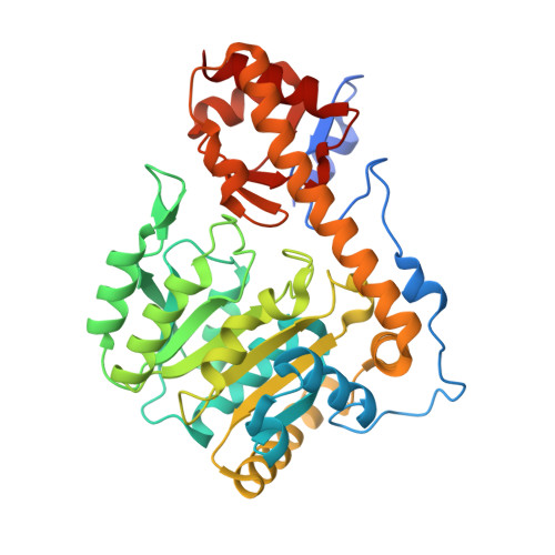
    The structures of three aspartate aminotransferases (AATs) from eukaryotic pathogens were solved within the Seattle Structural Genomics Center for Infectious Disease (SSGCID). Both the open and closed conformations of AAT were observed. Pyridoxal phosphate was bound to the active site via a Schiff base to a conserved lysine. An active-site mutant showed that Trypanosoma brucei AAT still binds pyridoxal phosphate even in the absence of the tethering lysine. The structures highlight the challenges for the structure-based design of inhibitors targeting the active site, while showing options for inhibitor design targeting the N-terminal arm.


  • Organizational Affiliation
    • Seattle Structural Genomics Center for Infectious Disease, http://www.ssgcid.org, USA.

Macromolecule Content 

  • Total Structure Weight: 88.52 kDa 
  • Atom Count: 6,708 
  • Modeled Residue Count: 760 
  • Deposited Residue Count: 792 
  • Unique protein chains: 1

Macromolecules

Find similar proteins by:|  3D Structure
Entity ID: 1
MoleculeChains  Sequence LengthOrganismDetailsImage
Aspartate aminotransferase, mitochondrial
A, B
396Trypanosoma brucei brucei TREU927Mutation(s): 1 
Gene Names: Tb11.02.2740
EC: 2.6.1.1
UniProt
Find proteins for Q385Q9 (Trypanosoma brucei brucei (strain 927/4 GUTat10.1))
Explore Q385Q9 
Go to UniProtKB:  Q385Q9
Entity Groups
Sequence Clusters30% Identity50% Identity70% Identity90% Identity95% Identity100% Identity
UniProt GroupQ385Q9
Sequence Annotations
Expand
Reference Sequence

Experimental Data & Validation

Experimental Data

  • Method: X-RAY DIFFRACTION
  • Resolution: 1.70 Å
  • R-Value Free:  0.183 (Depositor), 0.191 (DCC) 
  • R-Value Work:  0.153 (Depositor) 
  • R-Value Observed: 0.154 (Depositor) 
Space Group: P 1 21 1
Unit Cell:
Length ( Å )Angle ( ˚ )
a = 62.39α = 90
b = 96.66β = 111.03
c = 81.61γ = 90
Software Package:
Software NamePurpose
XDSdata reduction
XSCALEdata scaling
PHENIXrefinement
Cootmodel building
PDB_EXTRACTdata extraction

Structure Validation

View Full Validation Report


Ligand Structure Quality Assessment 


Entry History 

Deposition Data

Revision History  (Full details and data files)

  • Version 1.0: 2014-09-24
    Type: Initial release
  • Version 1.1: 2015-05-06
    Changes: Database references
  • Version 1.2: 2015-05-20
    Changes: Database references
  • Version 1.3: 2017-11-22
    Changes: Advisory, Derived calculations, Other, Refinement description, Source and taxonomy, Structure summary
  • Version 1.4: 2023-09-27
    Changes: Data collection, Database references, Refinement description
  • Version 2.0: 2024-12-25
    Changes: Advisory, Atomic model, Data collection, Derived calculations, Structure summary