4V4E | pdb_00004v4e

Crystal Structure of Pyrogallol-Phloroglucinol Transhydroxylase from Pelobacter acidigallici complexed with inhibitor 1,2,4,5-tetrahydroxy-benzene


Experimental Data Snapshot

  • Method: X-RAY DIFFRACTION
  • Resolution: 2.00 Å
  • R-Value Free: 
    0.203 (Depositor) 
  • R-Value Work: 
    0.172 (Depositor) 
  • R-Value Observed: 
    0.172 (Depositor) 

Starting Model: experimental
View more details

wwPDB Validation 3D Report Full Report

Validation slider image for 4V4E

This is version 1.2 of the entry. See complete history

Literature

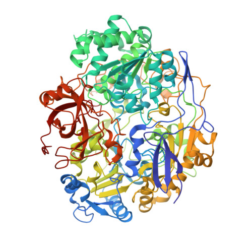
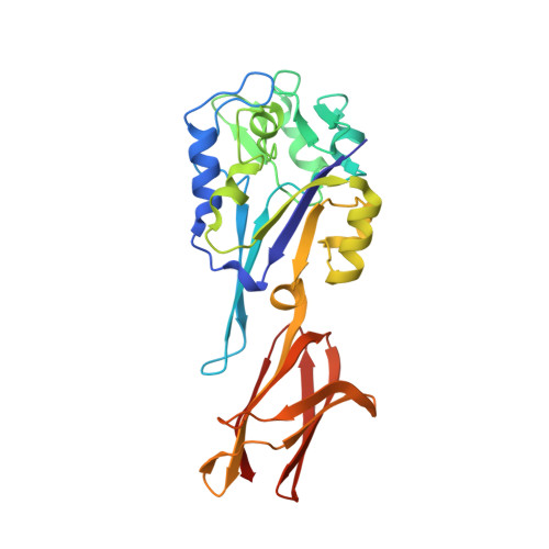
Crystal structure of pyrogallol-phloroglucinol transhydroxylase, an Mo enzyme capable of intermolecular hydroxyl transfer between phenols

Messerschmidt, A.Niessen, H.Abt, D.Einsle, O.Schink, B.Kroneck, P.M.H.

(2004) Proc Natl Acad Sci U S A 101: 11571-11576

  • DOI: https://doi.org/10.1073/pnas.0404378101
  • Primary Citation Related Structures: 
    4V4C, 4V4D, 4V4E

  • PubMed Abstract: 

    The Mo enzyme transhydroxylase from the anaerobic microorganism Pelobacter acidigallici catalyzes the conversion of pyrogallol to phloroglucinol. Such trihydroxybenzenes and their derivatives represent important building blocks of plant polymers. None of the transferred hydroxyl groups originates from water during transhydroxylation; instead a cosubstrate, such as 1,2,3,5-tetrahydroxybenzene, is used in a reaction without apparent electron transfer. Here, we report on the crystal structure of the enzyme in the reduced Mo(IV) state, which we solved by single anomalous-diffraction technique. It represents the largest structure (1,149 amino acid residues per molecule, 12 independent molecules per unit cell), which has been solved so far by single anomalous-diffraction technique. Tranhydroxylase is a heterodimer, with the active Mo-molybdopterin guanine dinucleotide (MGD)(2) site in the alpha-subunit, and three [4Fe-4S] centers in the beta-subunit. The latter subunit carries a seven-stranded, mainly antiparallel beta-barrel domain. We propose a scheme for the transhydroxylation reaction based on 3D structures of complexes of the enzyme with various polyphenols serving either as substrate or inhibitor.


  • Organizational Affiliation
    • Abteilung Strukturforschung, Max-Planck-Institut für Biochemie, Am Klopferspitz 18, 82152 Martinsried, Germany.

Macromolecule Content 

  • Total Structure Weight: 1,601.92 kDa 
  • Atom Count: 121,823 
  • Modeled Residue Count: 13,788 
  • Deposited Residue Count: 13,788 
  • Unique protein chains: 2

Macromolecules

Find similar proteins by:|  3D Structure
Entity ID: 1
MoleculeChains  Sequence LengthOrganismDetailsImage
Pyrogallol hydroxytransferase large subunit
A, C, E, G, I
A, C, E, G, I, K, M, O, Q, S, U, W
875Pelobacter acidigalliciMutation(s): 0 
EC: 1.97.1.2
UniProt
Find proteins for P80563 (Pelobacter acidigallici)
Explore P80563 
Go to UniProtKB:  P80563
Entity Groups
Sequence Clusters30% Identity50% Identity70% Identity90% Identity95% Identity100% Identity
UniProt GroupP80563
Sequence Annotations
Expand
Reference Sequence
Find similar proteins by:|  3D Structure
Entity ID: 2
MoleculeChains  Sequence LengthOrganismDetailsImage
Pyrogallol hydroxytransferase small subunit
B, D, F, H, J
B, D, F, H, J, L, N, P, R, T, V, X
274Pelobacter acidigalliciMutation(s): 0 
EC: 1.97.1.2
UniProt
Find proteins for P80564 (Pelobacter acidigallici)
Explore P80564 
Go to UniProtKB:  P80564
Entity Groups
Sequence Clusters30% Identity50% Identity70% Identity90% Identity95% Identity100% Identity
UniProt GroupP80564
Sequence Annotations
Expand
Reference Sequence

Small Molecules

Ligands 5 Unique
IDChains Name / Formula / InChI Key2D Diagram3D Interactions
MGD

Query on MGD


Download:Ideal Coordinates CCD File
AA [auth A]
AB [auth G]
BB [auth G]
BC [auth M]
CC [auth M]
AA [auth A],
AB [auth G],
BB [auth G],
BC [auth M],
CC [auth M],
CD [auth S],
DD [auth S],
IA [auth C],
JA [auth C],
JB [auth I],
KB [auth I],
KC [auth O],
LC [auth O],
LD [auth U],
MD [auth U],
RA [auth E],
SA [auth E],
SB [auth K],
TB [auth K],
TC [auth Q],
UC [auth Q],
UD [auth W],
VD [auth W],
Z [auth A]
2-AMINO-5,6-DIMERCAPTO-7-METHYL-3,7,8A,9-TETRAHYDRO-8-OXA-1,3,9,10-TETRAAZA-ANTHRACEN-4-ONE GUANOSINE DINUCLEOTIDE
C20 H26 N10 O13 P2 S2
VQAGYJCYOLHZDH-ILXWUORBSA-N
SF4

Query on SF4


Download:Ideal Coordinates CCD File
AD [auth R]
AE [auth X]
BE [auth X]
EA [auth B]
FA [auth B]
AD [auth R],
AE [auth X],
BE [auth X],
EA [auth B],
FA [auth B],
FB [auth H],
GA [auth B],
GB [auth H],
GC [auth N],
HB [auth H],
HC [auth N],
HD [auth T],
IC [auth N],
ID [auth T],
JD [auth T],
NA [auth D],
OA [auth D],
OB [auth J],
PA [auth D],
PB [auth J],
PC [auth P],
QB [auth J],
QC [auth P],
QD [auth V],
RC [auth P],
RD [auth V],
SD [auth V],
WA [auth F],
XA [auth F],
XB [auth L],
YA [auth F],
YB [auth L],
YC [auth R],
ZB [auth L],
ZC [auth R],
ZD [auth X]
IRON/SULFUR CLUSTER
Fe4 S4
LJBDFODJNLIPKO-UHFFFAOYSA-N
BTT

Query on BTT


Download:Ideal Coordinates CCD File
CA [auth A]
DB [auth G]
EC [auth M]
FD [auth S]
LA [auth C]
CA [auth A],
DB [auth G],
EC [auth M],
FD [auth S],
LA [auth C],
MB [auth I],
NC [auth O],
OD [auth U],
UA [auth E],
VB [auth K],
WC [auth Q],
XD [auth W]
BENZENE-1,2,4,5-TETROL
C6 H6 O4
UYQMSQMCIYSXOW-UHFFFAOYSA-N
4MO

Query on 4MO


Download:Ideal Coordinates CCD File
BA [auth A]
CB [auth G]
DC [auth M]
ED [auth S]
KA [auth C]
BA [auth A],
CB [auth G],
DC [auth M],
ED [auth S],
KA [auth C],
LB [auth I],
MC [auth O],
ND [auth U],
TA [auth E],
UB [auth K],
VC [auth Q],
WD [auth W]
MOLYBDENUM(IV) ION
Mo
ZIKKVZAYJJZBGE-UHFFFAOYSA-N
CA

Query on CA


Download:Ideal Coordinates CCD File
AC [auth M]
BD [auth S]
DA [auth B]
EB [auth H]
FC [auth N]
AC [auth M],
BD [auth S],
DA [auth B],
EB [auth H],
FC [auth N],
GD [auth T],
HA [auth C],
IB [auth I],
JC [auth O],
KD [auth U],
MA [auth D],
NB [auth J],
OC [auth P],
PD [auth V],
QA [auth E],
RB [auth K],
SC [auth Q],
TD [auth W],
VA [auth F],
WB [auth L],
XC [auth R],
Y [auth A],
YD [auth X],
ZA [auth G]
CALCIUM ION
Ca
BHPQYMZQTOCNFJ-UHFFFAOYSA-N

Experimental Data & Validation

Experimental Data

  • Method: X-RAY DIFFRACTION
  • Resolution: 2.00 Å
  • R-Value Free:  0.203 (Depositor) 
  • R-Value Work:  0.172 (Depositor) 
  • R-Value Observed: 0.172 (Depositor) 
Space Group: P 1
Unit Cell:
Length ( Å )Angle ( ˚ )
a = 172.575α = 63.83
b = 178.437β = 64.4
c = 179.665γ = 65.04
Software Package:
Software NamePurpose
CNSrefinement
MOSFLMdata reduction
CCP4data scaling
CNSphasing

Structure Validation

View Full Validation Report


Entry History 

Deposition Data

Revision History  (Full details and data files)

  • Version 1.0: 2014-07-09
    Type: Initial release
  • Version 1.1: 2014-12-10
    Changes: Other
  • Version 1.2: 2023-09-20
    Changes: Advisory, Data collection, Database references, Derived calculations, Refinement description