4V3B | pdb_00004v3b

The structure of alpha2,3-sialyltransferase variant 1 from Pasteurella dagmatis in complex with the donor product CMP


Experimental Data Snapshot

  • Method: X-RAY DIFFRACTION
  • Resolution: 2.18 Å
  • R-Value Free: 
    0.238 (Depositor), 0.244 (DCC) 
  • R-Value Work: 
    0.186 (Depositor), 0.196 (DCC) 
  • R-Value Observed: 
    0.189 (Depositor) 

Starting Model: experimental
View more details

wwPDB Validation 3D Report Full Report

Validation slider image for 4V3B

Ligand Structure Quality Assessment 


This is version 2.0 of the entry. See complete history

Literature

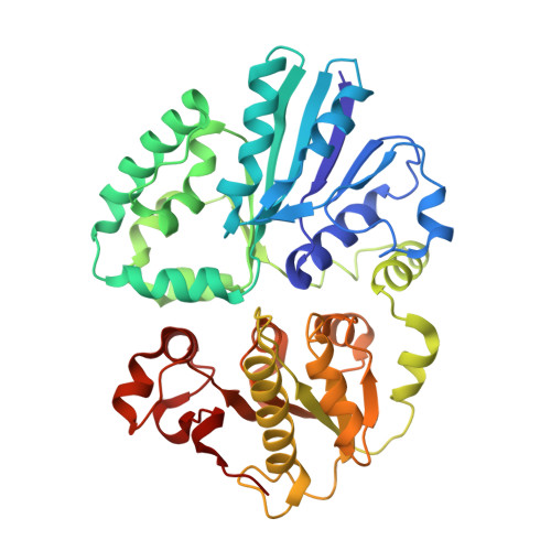
Complete Switch from Alpha2,3- to Alpha2,6-Regioselectivity in Pasteurella Dagmatis Beta-D-Galactoside Sialyltransferase by Active-Site Redesign

Schmoelzer, K.Czabany, T.Pavkov-Keller, T.Luley-Goedl, C.Ribitsch, D.Schwab, H.Gruber, K.Nidetzky, B.

(2015) Chem Commun (Camb) 51: 3083

  • DOI: https://doi.org/10.1039/c4cc09772f
  • Primary Citation Related Structures: 
    4V2U, 4V38, 4V39, 4V3B, 4V3C

  • PubMed Abstract: 

    Structure-guided active-site redesign of a family GT-80 β-D-galactoside sialyltransferase (from Pasteurella dagmatis) to change enzyme regioselectivity from α-2,3 in the wild type to α-2,6 in a P7H-M117A double mutant is reported. Biochemical data for sialylation of lactose together with protein crystal structures demonstrate highly precise enzyme engineering.


  • Organizational Affiliation
    • Austrian Centre of Industrial Biotechnology, Petersgasse 14, 8010 Graz, Austria.

Macromolecule Content 

  • Total Structure Weight: 46.03 kDa 
  • Atom Count: 3,512 
  • Modeled Residue Count: 388 
  • Deposited Residue Count: 388 
  • Unique protein chains: 1

Macromolecules

Find similar proteins by:|  3D Structure
Entity ID: 1
MoleculeChains  Sequence LengthOrganismDetailsImage
SIALYLTRANSFERASE388Pasteurella dagmatisMutation(s): 1 
UniProt
Find proteins for K9UUI6 (Pasteurella dagmatis)
Explore K9UUI6 
Go to UniProtKB:  K9UUI6
Entity Groups
Sequence Clusters30% Identity50% Identity70% Identity90% Identity95% Identity100% Identity
UniProt GroupK9UUI6
Sequence Annotations
Expand
Reference Sequence

Small Molecules

Ligands 1 Unique
IDChains Name / Formula / InChI Key2D Diagram3D Interactions
C5P

Query on C5P


Download:Ideal Coordinates CCD File
B [auth A]CYTIDINE-5'-MONOPHOSPHATE
C9 H14 N3 O8 P
IERHLVCPSMICTF-XVFCMESISA-N

Experimental Data & Validation

Experimental Data

  • Method: X-RAY DIFFRACTION
  • Resolution: 2.18 Å
  • R-Value Free:  0.238 (Depositor), 0.244 (DCC) 
  • R-Value Work:  0.186 (Depositor), 0.196 (DCC) 
  • R-Value Observed: 0.189 (Depositor) 
Space Group: P 65
Unit Cell:
Length ( Å )Angle ( ˚ )
a = 113.555α = 90
b = 113.555β = 90
c = 65.74γ = 120
Software Package:
Software NamePurpose
REFMACrefinement
XDSdata reduction
XDSdata scaling
PHASERphasing

Structure Validation

View Full Validation Report


Ligand Structure Quality Assessment 


Entry History 

Deposition Data

Revision History  (Full details and data files)

  • Version 1.0: 2015-04-08
    Type: Initial release
  • Version 1.1: 2024-01-10
    Changes: Data collection, Database references, Derived calculations, Other, Refinement description
  • Version 2.0: 2025-12-17
    Changes: Atomic model, Data collection, Derived calculations, Non-polymer description, Structure summary