4V2P | pdb_00004v2p

Ketosynthase MxnB


Experimental Data Snapshot

  • Method: X-RAY DIFFRACTION
  • Resolution: 1.67 Å
  • R-Value Free: 
    0.177 (Depositor), 0.185 (DCC) 
  • R-Value Work: 
    0.146 (Depositor), 0.159 (DCC) 
  • R-Value Observed: 
    0.147 (Depositor) 

wwPDB Validation 3D Report Full Report

Validation slider image for 4V2P

This is version 1.3 of the entry. See complete history

Literature

In vitro reconstitution of alpha-pyrone ring formation in myxopyronin biosynthesis.

Sucipto, H.Sahner, J.H.Prusov, E.Wenzel, S.C.Hartmann, R.W.Koehnke, J.Muller, R.

(2015) Chem Sci 6: 5076-5085

  • DOI: https://doi.org/10.1039/c5sc01013f
  • Primary Citation Related Structures: 
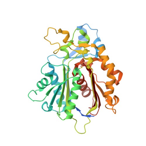
    4V2P

  • PubMed Abstract: 

    Myxopyronins are α-pyrone antibiotics produced by the terrestrial bacterium Myxococcus fulvus Mx f50 and possess antibacterial activity against Gram-positive and Gram-negative pathogens. They target the bacterial RNA polymerase (RNAP) "switch region" as non-competitive inhibitors and display no cross-resistance to the established RNAP inhibitor rifampicin. Recent analysis of the myxopyronin biosynthetic pathway led to the hypothesis that this secondary metabolite is produced from two separate polyketide parts, which are condensed by the stand-alone ketosynthase MxnB. Using in vitro assays we show that MxnB catalyzes a unique condensation reaction forming the α-pyrone ring of myxopyronins from two activated acyl chains in form of their β-keto intermediates. MxnB is able to accept thioester substrates coupled to either N -acetylcysteamine (NAC) or a specific carrier protein (CP). The turnover rate of MxnB for substrates bound to CP was 12-fold higher than for NAC substrates, demonstrating the importance of protein-protein interactions in polyketide synthase (PKS) systems. The crystal structure of MxnB reveals the enzyme to be an unusual member of the ketosynthase group capable of binding and condensing two long alkyl chains bound to carrier proteins. The geometry of the two binding tunnels supports the biochemical data and allows us to propose an order of reaction, which is supported by the identification of novel myxopyronin congeners in the extract of the producer strain. Insights into the mechanism of this unique condensation reaction do not only expand our knowledge regarding the thiolase enzyme family but also opens up opportunities for PKS bioengineering to achieve directed structural modifications.


  • Organizational Affiliation
    • Department of Microbial Natural Products , Helmholtz Institute for Pharmaceutical Research Saarland , Building C2 3 , 66123 Saarbrücken , Germany . Email: rolf.mueller@helmholtz-hzi.de.

Macromolecule Content 

  • Total Structure Weight: 78.91 kDa 
  • Atom Count: 5,884 
  • Modeled Residue Count: 657 
  • Deposited Residue Count: 702 
  • Unique protein chains: 1

Macromolecules

Find similar proteins by:|  3D Structure
Entity ID: 1
MoleculeChains  Sequence LengthOrganismDetailsImage
KETOSYNTHASE
A, B
351Myxococcus fulvusMutation(s): 0 
UniProt
Find proteins for T1SF45 (Myxococcus fulvus)
Explore T1SF45 
Go to UniProtKB:  T1SF45
Entity Groups
Sequence Clusters30% Identity50% Identity70% Identity90% Identity95% Identity100% Identity
UniProt GroupT1SF45
Sequence Annotations
Expand
Reference Sequence

Small Molecules

Ligands 4 Unique
IDChains Name / Formula / InChI Key2D Diagram3D Interactions
MPD

Query on MPD


Download:Ideal Coordinates CCD File
F [auth A],
G [auth A],
N [auth B],
O [auth B]
(4S)-2-METHYL-2,4-PENTANEDIOL
C6 H14 O2
SVTBMSDMJJWYQN-YFKPBYRVSA-N
PO4

Query on PO4


Download:Ideal Coordinates CCD File
C [auth A]
D [auth A]
E [auth A]
J [auth B]
K [auth B]
C [auth A],
D [auth A],
E [auth A],
J [auth B],
K [auth B],
L [auth B],
M [auth B]
PHOSPHATE ION
O4 P
NBIIXXVUZAFLBC-UHFFFAOYSA-K
CL

Query on CL


Download:Ideal Coordinates CCD File
H [auth A]CHLORIDE ION
Cl
VEXZGXHMUGYJMC-UHFFFAOYSA-M
NA

Query on NA


Download:Ideal Coordinates CCD File
I [auth A],
P [auth B]
SODIUM ION
Na
FKNQFGJONOIPTF-UHFFFAOYSA-N
Modified Residues  1 Unique
IDChains TypeFormula2D DiagramParent
CSD
Query on CSD
A, B
L-PEPTIDE LINKINGC3 H7 N O4 SCYS

Experimental Data & Validation

Experimental Data

  • Method: X-RAY DIFFRACTION
  • Resolution: 1.67 Å
  • R-Value Free:  0.177 (Depositor), 0.185 (DCC) 
  • R-Value Work:  0.146 (Depositor), 0.159 (DCC) 
  • R-Value Observed: 0.147 (Depositor) 
Space Group: P 1
Unit Cell:
Length ( Å )Angle ( ˚ )
a = 50.19α = 104.75
b = 58.79β = 105.48
c = 62.82γ = 93.9
Software Package:
Software NamePurpose
REFMACrefinement
PHASERphasing

Structure Validation

View Full Validation Report


Entry History 

Deposition Data

  • Released Date: 2015-08-26 
  • Deposition Author(s): Koehnke, J.

Revision History  (Full details and data files)

  • Version 1.0: 2015-08-26
    Type: Initial release
  • Version 1.1: 2017-09-27
    Changes: Data collection
  • Version 1.2: 2018-04-18
    Changes: Data collection, Database references
  • Version 1.3: 2024-10-16
    Changes: Data collection, Database references, Derived calculations, Other, Structure summary