4V2C

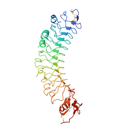
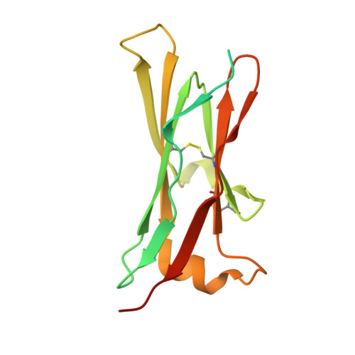
mouse FLRT2 LRR domain in complex with rat Unc5D Ig1 domain


Experimental Data Snapshot

  • Method: X-RAY DIFFRACTION
  • Resolution: 4.00 Å
  • R-Value Free: 0.335 
  • R-Value Work: 0.318 
  • R-Value Observed: 0.319 

Starting Model: experimental
View more details

wwPDB Validation   3D Report Full Report


This is version 1.2 of the entry. See complete history


Literature

Flrt Structure: Balancing Repulsion and Cell Adhesion in Cortical and Vascular Development

Seiradake, E.Del Toro, D.Nagel, D.Cop, F.Haertl, R.Ruff, T.Seyit-Bremer, G.Harlos, K.Border, E.C.Acker-Palmer, A.Jones, E.Y.Klein, R.

(2014) Neuron 84: 370

  • DOI: https://doi.org/10.1016/j.neuron.2014.10.008
  • Primary Citation of Related Structures:  
    4V2A, 4V2B, 4V2C, 4V2D, 4V2E

  • PubMed Abstract: 

    FLRTs are broadly expressed proteins with the unique property of acting as homophilic cell adhesion molecules and as heterophilic repulsive ligands of Unc5/Netrin receptors. How these functions direct cell behavior and the molecular mechanisms involved remain largely unclear. Here we use X-ray crystallography to reveal the distinct structural bases for FLRT-mediated cell adhesion and repulsion in neurons. We apply this knowledge to elucidate FLRT functions during cortical development. We show that FLRTs regulate both the radial migration of pyramidal neurons, as well as their tangential spread. Mechanistically, radial migration is controlled by repulsive FLRT2-Unc5D interactions, while spatial organization in the tangential axis involves adhesive FLRT-FLRT interactions. Further, we show that the fundamental mechanisms of FLRT adhesion and repulsion are conserved between neurons and vascular endothelial cells. Our results reveal FLRTs as powerful guidance factors with structurally encoded repulsive and adhesive surfaces.


  • Organizational Affiliation

    Division of Structural Biology, Wellcome Trust Centre for Human Genetics, University of Oxford, Roosevelt Drive, OX3 7BN Oxford, UK.


Macromolecules
Find similar proteins by:  (by identity cutoff)  |  3D Structure
Entity ID: 1
MoleculeChains Sequence LengthOrganismDetailsImage
FIBRONECTIN LEUCINE RICH TRANSMEMBRANE PROTEIN 2
A, C
328Mus musculusMutation(s): 0 
UniProt
Find proteins for Q8BLU0 (Mus musculus)
Go to UniProtKB:  Q8BLU0
Sequence Annotations
Expand
  • Reference Sequence
Find similar proteins by:  (by identity cutoff)  |  3D Structure
Entity ID: 2
MoleculeChains Sequence LengthOrganismDetailsImage
PROTEIN UNC5D
B, D
161Rattus norvegicusMutation(s): 0 
UniProt
Find proteins for F1LW30 (Rattus norvegicus)
Go to UniProtKB:  F1LW30
Sequence Annotations
Expand
  • Reference Sequence
Experimental Data & Validation

Experimental Data

  • Method: X-RAY DIFFRACTION
  • Resolution: 4.00 Å
  • R-Value Free: 0.335 
  • R-Value Work: 0.318 
  • R-Value Observed: 0.319 
  • Space Group: C 1 2 1
Unit Cell:
Length ( Å )Angle ( ˚ )
a = 180.14α = 90
b = 150.91β = 102.79
c = 67.22γ = 90
Software Package:
Software NamePurpose
BUSTERrefinement
xia2data reduction
XDSdata reduction
xia2data scaling
XSCALEdata scaling
PHASERphasing

Structure Validation

View Full Validation Report


Entry History 

Revision History  (Full details and data files)

  • Version 1.0: 2014-11-05
    Type: Initial release
  • Version 1.1: 2015-07-22
    Changes: Database references
  • Version 1.2: 2024-01-10
    Changes: Data collection, Database references, Other, Refinement description