4V0H | pdb_00004v0h

Human metallo beta lactamase domain containing protein 1 (hMBLAC1)


Experimental Data Snapshot

  • Method: X-RAY DIFFRACTION
  • Resolution: 1.79 Å
  • R-Value Free: 
    0.211 (Depositor), 0.220 (DCC) 
  • R-Value Work: 
    0.182 (Depositor), 0.193 (DCC) 
  • R-Value Observed: 
    0.184 (Depositor) 

wwPDB Validation 3D Report Full Report

Validation slider image for 4V0H

This is version 1.3 of the entry. See complete history

Literature

Biosynthesis of histone messenger RNA employs a specific 3' end endonuclease.

Pettinati, I.Grzechnik, P.Ribeiro de Almeida, C.Brem, J.McDonough, M.A.Dhir, S.Proudfoot, N.J.Schofield, C.J.

(2018) Elife 7

  • DOI: https://doi.org/10.7554/eLife.39865
  • Primary Citation Related Structures: 
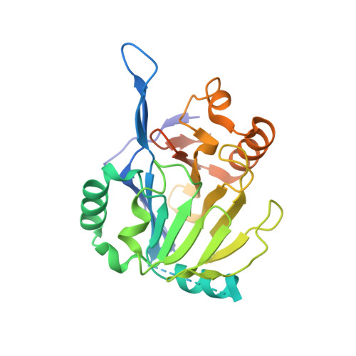
    4V0H

  • PubMed Abstract: 

    Replication-dependent (RD) core histone mRNA produced during S-phase is the only known metazoan protein-coding mRNA presenting a 3' stem-loop instead of the otherwise universal polyA tail. A metallo β-lactamase (MBL) fold enzyme, cleavage and polyadenylation specificity factor 73 (CPSF73), is proposed to be the sole endonuclease responsible for 3' end processing of both mRNA classes. We report cellular, genetic, biochemical, substrate selectivity, and crystallographic studies providing evidence that an additional endoribonuclease, MBL domain containing protein 1 (MBLAC1), is selective for 3' processing of RD histone pre-mRNA during the S-phase of the cell cycle. Depletion of MBLAC1 in cells significantly affects cell cycle progression thus identifying MBLAC1 as a new type of S-phase-specific cancer target.


  • Organizational Affiliation
    • Department of Chemistry, University of Oxford, Oxford, United Kingdom.

Macromolecule Content 

  • Total Structure Weight: 109.64 kDa 
  • Atom Count: 6,787 
  • Modeled Residue Count: 885 
  • Deposited Residue Count: 1,064 
  • Unique protein chains: 1

Macromolecules

Find similar proteins by:|  3D Structure
Entity ID: 1
MoleculeChains  Sequence LengthOrganismDetailsImage
METALLO-BETA-LACTAMASE DOMAIN-CONTAINING PROTEIN 1 1
A, B, C, D
266Homo sapiensMutation(s): 0 
EC: 3 (PDB Primary Data), 3.1.27 (UniProt)
UniProt & NIH Common Fund Data Resources
Find proteins for A4D2B0 (Homo sapiens)
Explore A4D2B0 
Go to UniProtKB:  A4D2B0
PHAROS:  A4D2B0
GTEx:  ENSG00000214309 
Entity Groups
Sequence Clusters30% Identity50% Identity70% Identity90% Identity95% Identity100% Identity
UniProt GroupA4D2B0
Sequence Annotations
Expand
Reference Sequence

Small Molecules

Ligands 2 Unique
IDChains Name / Formula / InChI Key2D Diagram3D Interactions
GOL

Query on GOL


Download:Ideal Coordinates CCD File
I [auth B],
L [auth C],
O [auth D]
GLYCEROL
C3 H8 O3
PEDCQBHIVMGVHV-UHFFFAOYSA-N
FE

Query on FE


Download:Ideal Coordinates CCD File
E [auth A]
F [auth A]
G [auth B]
H [auth B]
J [auth C]
E [auth A],
F [auth A],
G [auth B],
H [auth B],
J [auth C],
K [auth C],
M [auth D],
N [auth D]
FE (III) ION
Fe
VTLYFUHAOXGGBS-UHFFFAOYSA-N

Experimental Data & Validation

Experimental Data

  • Method: X-RAY DIFFRACTION
  • Resolution: 1.79 Å
  • R-Value Free:  0.211 (Depositor), 0.220 (DCC) 
  • R-Value Work:  0.182 (Depositor), 0.193 (DCC) 
  • R-Value Observed: 0.184 (Depositor) 
Space Group: P 1
Unit Cell:
Length ( Å )Angle ( ˚ )
a = 62.95α = 109.31
b = 67.13β = 105.4
c = 67.9γ = 90.17
Software Package:
Software NamePurpose
PHENIXrefinement
XDSdata reduction
xia2data scaling
PHASERphasing

Structure Validation

View Full Validation Report


Entry History 

Deposition Data

Revision History  (Full details and data files)

  • Version 1.0: 2016-01-13
    Type: Initial release
  • Version 1.1: 2018-02-21
    Changes: Database references
  • Version 1.2: 2019-11-20
    Changes: Database references, Other
  • Version 1.3: 2024-05-08
    Changes: Data collection, Database references, Derived calculations