4USJ | pdb_00004usj

N-acetylglutamate kinase from Arabidopsis thaliana in complex with PII from Chlamydomonas reinhardtii


Experimental Data Snapshot

  • Method: X-RAY DIFFRACTION
  • Resolution: 2.85 Å
  • R-Value Free: 
    0.220 (Depositor), 0.207 (DCC) 
  • R-Value Work: 
    0.189 (Depositor), 0.173 (DCC) 
  • R-Value Observed: 
    0.190 (Depositor) 

Starting Model: experimental
View more details

wwPDB Validation 3D Report Full Report

Validation slider image for 4USJ

Ligand Structure Quality Assessment 


This is version 1.1 of the entry. See complete history

Literature

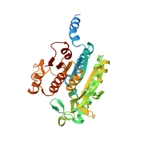
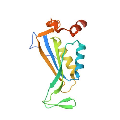
A Widespread Glutamine-Sensing Mechanism in the Plant Kingdom.

Chellamuthu, V.R.Ermilova, E.Lapina, T.Luddecke, J.Minaeva, E.Herrmann, C.Hartmann, M.D.Forchhammer, K.

(2014) Cell 159: 1188

  • DOI: https://doi.org/10.1016/j.cell.2014.10.015
  • Primary Citation Related Structures: 
    4USH, 4USI, 4USJ

  • PubMed Abstract: 

    Glutamine is the primary metabolite of nitrogen assimilation from inorganic nitrogen sources in microorganisms and plants. The ability to monitor cellular nitrogen status is pivotal for maintaining metabolic homeostasis and sustaining growth. The present study identifies a glutamine-sensing mechanism common in the entire plant kingdom except Brassicaceae. The plastid-localized PII signaling protein controls, in a glutamine-dependent manner, the key enzyme of the ornithine synthesis pathway, N-acetyl-l-glutamate kinase (NAGK), that leads to arginine and polyamine formation. Crystal structures reveal that the plant-specific C-terminal extension of PII, which we term the Q loop, forms a low-affinity glutamine-binding site. Glutamine binding alters PII conformation, promoting interaction and activation of NAGK. The binding motif is highly conserved in plants except Brassicaceae. A functional Q loop restores glutamine sensing in a recombinant Arabidopsis thaliana PII protein, demonstrating the modular concept of the glutamine-sensing mechanism adopted by PII proteins during the evolution of plant chloroplasts.


  • Organizational Affiliation
    • Interfaculty Institute for Microbiology and Infection Medicine, University of Tübingen, Auf der Morgenstelle 28, 72076 Tübingen, Germany; Department of Protein Evolution, Max Planck Institute for Developmental Biology, Spemannstrasse 35, 72076 Tübingen, Germany.

Macromolecule Content 

  • Total Structure Weight: 103.61 kDa 
  • Atom Count: 6,567 
  • Modeled Residue Count: 846 
  • Deposited Residue Count: 944 
  • Unique protein chains: 2

Macromolecules

Find similar proteins by:|  3D Structure
Entity ID: 1
MoleculeChains  Sequence LengthOrganismDetailsImage
ACETYLGLUTAMATE KINASE, CHLOROPLASTIC
A, B
318Arabidopsis thalianaMutation(s): 0 
EC: 2.7.2.8
UniProt
Find proteins for Q9SCL7 (Arabidopsis thaliana)
Explore Q9SCL7 
Go to UniProtKB:  Q9SCL7
Entity Groups
Sequence Clusters30% Identity50% Identity70% Identity90% Identity95% Identity100% Identity
UniProt GroupQ9SCL7
Sequence Annotations
Expand
Reference Sequence
Find similar proteins by:|  3D Structure
Entity ID: 2
MoleculeChains  Sequence LengthOrganismDetailsImage
NITROGEN REGULATORY PROTEIN PII
C, D
154Chlamydomonas reinhardtiiMutation(s): 0 

Small Molecules

Ligands 7 Unique
IDChains Name / Formula / InChI Key2D Diagram3D Interactions
ATP

Query on ATP


Download:Ideal Coordinates CCD File
K [auth C],
N [auth D]
ADENOSINE-5'-TRIPHOSPHATE
C10 H16 N5 O13 P3
ZKHQWZAMYRWXGA-KQYNXXCUSA-N
ADP

Query on ADP


Download:Ideal Coordinates CCD File
F [auth A]ADENOSINE-5'-DIPHOSPHATE
C10 H15 N5 O10 P2
XTWYTFMLZFPYCI-KQYNXXCUSA-N
X2W

Query on X2W


Download:Ideal Coordinates CCD File
J [auth B]N-ACETYL-L-GLUTAMYL 5-PHOSPHATE
C7 H12 N O8 P
FCVIHFVSXHOPSW-YFKPBYRVSA-N
NLG

Query on NLG


Download:Ideal Coordinates CCD File
G [auth A]N-ACETYL-L-GLUTAMATE
C7 H11 N O5
RFMMMVDNIPUKGG-YFKPBYRVSA-N
ARG

Query on ARG


Download:Ideal Coordinates CCD File
E [auth A],
I [auth B]
ARGININE
C6 H15 N4 O2
ODKSFYDXXFIFQN-BYPYZUCNSA-O
GLN

Query on GLN


Download:Ideal Coordinates CCD File
M [auth C],
P [auth D]
GLUTAMINE
C5 H10 N2 O3
ZDXPYRJPNDTMRX-VKHMYHEASA-N
MG

Query on MG


Download:Ideal Coordinates CCD File
H [auth A],
L [auth C],
O [auth D]
MAGNESIUM ION
Mg
JLVVSXFLKOJNIY-UHFFFAOYSA-N

Experimental Data & Validation

Experimental Data

  • Method: X-RAY DIFFRACTION
  • Resolution: 2.85 Å
  • R-Value Free:  0.220 (Depositor), 0.207 (DCC) 
  • R-Value Work:  0.189 (Depositor), 0.173 (DCC) 
  • R-Value Observed: 0.190 (Depositor) 
Space Group: P 21 3
Unit Cell:
Length ( Å )Angle ( ˚ )
a = 171.43α = 90
b = 171.43β = 90
c = 171.43γ = 90
Software Package:
Software NamePurpose
REFMACrefinement
XDSdata reduction
XDSdata scaling
MOLREPphasing

Structure Validation

View Full Validation Report


Ligand Structure Quality Assessment 


Entry History 

Deposition Data

Revision History  (Full details and data files)

  • Version 1.0: 2014-12-03
    Type: Initial release
  • Version 1.1: 2024-01-10
    Changes: Data collection, Database references, Derived calculations, Other, Refinement description