4UR6 | pdb_00004ur6

Structure of the type III fish antifreeze protein from Zoarces viviparus ZvAFP6


Experimental Data Snapshot

  • Method: X-RAY DIFFRACTION
  • Resolution: 1.20 Å
  • R-Value Free: 
    0.195 (Depositor), 0.190 (DCC) 
  • R-Value Work: 
    0.179 (Depositor), 0.172 (DCC) 
  • R-Value Observed: 
    0.180 (Depositor) 

Starting Model: experimental
View more details

wwPDB Validation 3D Report Full Report

Validation slider image for 4UR6

This is version 1.3 of the entry. See complete history

Literature

Purification, Crystal Structure Determination and Functional Characterization of Type III Antifreeze Proteins from the European Eelpout Zoarces Viviparus.

Wilkens, C.Poulsen, J.N.Ramlov, H.Lo Leggio, L.

(2014) Cryobiology 69: 163

  • DOI: https://doi.org/10.1016/j.cryobiol.2014.07.003
  • Primary Citation Related Structures: 
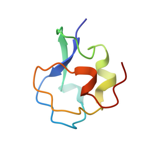
    4UR4, 4UR6

  • PubMed Abstract: 

    Antifreeze proteins (AFPs) are essential components of many organisms adaptation to cold temperatures. Fish type III AFPs are divided into two groups, SP isoforms being much less active than QAE1 isoforms. Two type III AFPs from Zoarces viviparus, a QAE1 (ZvAFP13) and an SP (ZvAFP6) isoform, are here characterized and their crystal structures determined. We conclude that the higher activity of the QAE1 isoforms cannot be attributed to single residues, but rather a combination of structural effects. Furthermore both ZvAFP6 and ZvAFP13 crystal structures have water molecules around T18 equivalent to the tetrahedral-like waters previously identified in a neutron crystal structure. Interestingly, ZvAFP6 forms dimers in the crystal, with a significant dimer interface. The presence of ZvAFP6 dimers was confirmed in solution by native electrophoresis and gel filtration. To our knowledge this is the first report of dimerization of AFP type III proteins.


  • Organizational Affiliation
    • Department of Chemistry, University of Copenhagen, Universitetsparken 5, DK-2100 Copenhagen, Denmark. Electronic address: cwil@bio.dtu.dk.

Macromolecule Content 

  • Total Structure Weight: 13.94 kDa 
  • Atom Count: 1,183 
  • Modeled Residue Count: 126 
  • Deposited Residue Count: 132 
  • Unique protein chains: 1

Macromolecules

Find similar proteins by:|  3D Structure
Entity ID: 1
MoleculeChains  Sequence LengthOrganismDetailsImage
TYPE III ANTIFREEZE PROTEIN 6
A, B
66Zoarces viviparusMutation(s): 0 
UniProt
Find proteins for R9S083 (Zoarces viviparus)
Explore R9S083 
Go to UniProtKB:  R9S083
Entity Groups
Sequence Clusters30% Identity50% Identity70% Identity90% Identity95% Identity100% Identity
UniProt GroupR9S083
Sequence Annotations
Expand
Reference Sequence

Experimental Data & Validation

Experimental Data

  • Method: X-RAY DIFFRACTION
  • Resolution: 1.20 Å
  • R-Value Free:  0.195 (Depositor), 0.190 (DCC) 
  • R-Value Work:  0.179 (Depositor), 0.172 (DCC) 
  • R-Value Observed: 0.180 (Depositor) 
Space Group: C 2 2 21
Unit Cell:
Length ( Å )Angle ( ˚ )
a = 75.57α = 90
b = 108.17β = 90
c = 38.25γ = 90
Software Package:
Software NamePurpose
REFMACrefinement
XDSdata reduction
XSCALEdata scaling
MOLREPphasing

Structure Validation

View Full Validation Report


Entry History 

Deposition Data

Revision History  (Full details and data files)

  • Version 1.0: 2014-07-23
    Type: Initial release
  • Version 1.1: 2014-08-27
    Changes: Database references
  • Version 1.2: 2018-01-17
    Changes: Data collection
  • Version 1.3: 2024-01-10
    Changes: Data collection, Database references, Derived calculations, Other, Refinement description