4UP9

Crystal structure of Entamoeba histolytica lysyl-tRNA synthetase in complex with ATP


Experimental Data Snapshot

  • Method: X-RAY DIFFRACTION
  • Resolution: 3.05 Å
  • R-Value Free: 0.263 
  • R-Value Work: 0.216 
  • R-Value Observed: 0.219 

Starting Model: experimental
View more details

wwPDB Validation   3D Report Full Report


Ligand Structure Quality Assessment 


This is version 1.4 of the entry. See complete history


Literature

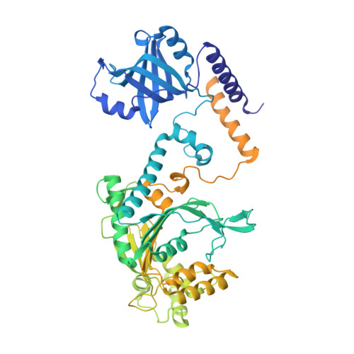
Crystal Structures of Entamoeba Histolytica Lysyl-tRNA Synthetase Reveal Conformational Changes Upon Lysine Binding and a Specific Helix Bundle Domain.

Bonnefond, L.Castro De Moura, M.Ribas De Pouplana, L.Nureki, O.

(2014) FEBS Lett 588: 4478

  • DOI: https://doi.org/10.1016/j.febslet.2014.10.019
  • Primary Citation of Related Structures:  
    4UP7, 4UP8, 4UP9, 4UPA

  • PubMed Abstract: 

    The class II lysyl-tRNA synthetases (KRS) are conserved aminoacyl-tRNA synthetases that attach lysine to the cognate tRNA in a two-step mechanism. The enzyme from the parasitic protozoan Entamoeba histolytica was crystallized in the presence of small ligands to generate snapshots of the lysine-adenylate formation. The residues involved in lysine activation are highly conserved and the active site closes around the lysyl-adenylate, as observed in bacterial KRS. The Entamoeba EMAPII-like polypeptide is not resolved in the crystals, but another Entamoeba-specific insertion could be modeled as a small helix bundle that may contribute to tRNA binding through interaction with the tRNA hinge.


  • Organizational Affiliation

    Department of Biophysics and Biochemistry, Graduate School of Science, The University of Tokyo, 2-11-16, Yayoi, Bunkyo, Tokyo 113-0032, Japan. lbonnefond@unistra.fr


Macromolecules
Find similar proteins by:  (by identity cutoff)  |  3D Structure
Entity ID: 1
MoleculeChains Sequence LengthOrganismDetailsImage
LYSINE--TRNA LIGASE777Entamoeba histolyticaMutation(s): 1 
EC: 6.1.1.6
UniProt
Find proteins for C4M7X2 (Entamoeba histolytica (strain ATCC 30459 / HM-1:IMSS / ABRM))
Explore C4M7X2 
Go to UniProtKB:  C4M7X2
Entity Groups  
Sequence Clusters30% Identity50% Identity70% Identity90% Identity95% Identity100% Identity
UniProt GroupC4M7X2
Sequence Annotations
Expand
  • Reference Sequence
Small Molecules
Ligands 1 Unique
IDChains Name / Formula / InChI Key2D Diagram3D Interactions
ATP
Query on ATP

Download Ideal Coordinates CCD File 
B [auth A]ADENOSINE-5'-TRIPHOSPHATE
C10 H16 N5 O13 P3
ZKHQWZAMYRWXGA-KQYNXXCUSA-N
Experimental Data & Validation

Experimental Data

  • Method: X-RAY DIFFRACTION
  • Resolution: 3.05 Å
  • R-Value Free: 0.263 
  • R-Value Work: 0.216 
  • R-Value Observed: 0.219 
  • Space Group: P 62
Unit Cell:
Length ( Å )Angle ( ˚ )
a = 155.551α = 90
b = 155.551β = 90
c = 92.926γ = 120
Software Package:
Software NamePurpose
PHENIXrefinement
XDSdata reduction
Aimlessdata scaling
PHASERphasing

Structure Validation

View Full Validation Report


Ligand Structure Quality Assessment 


Entry History 

Deposition Data

Revision History  (Full details and data files)

  • Version 1.0: 2014-10-29
    Type: Initial release
  • Version 1.1: 2014-11-05
    Changes: Database references
  • Version 1.2: 2014-11-26
    Changes: Database references
  • Version 1.3: 2014-12-10
    Changes: Database references
  • Version 1.4: 2024-01-10
    Changes: Data collection, Database references, Derived calculations, Other, Refinement description