4UJB

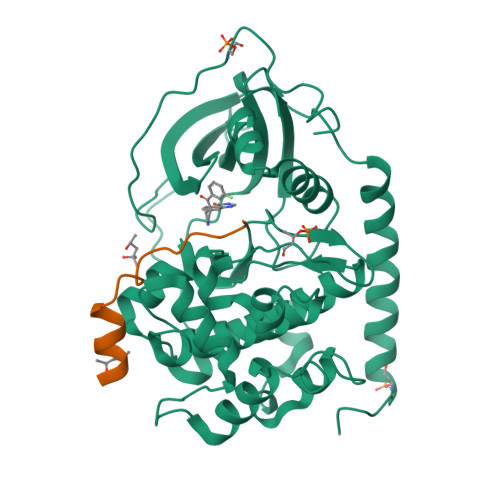
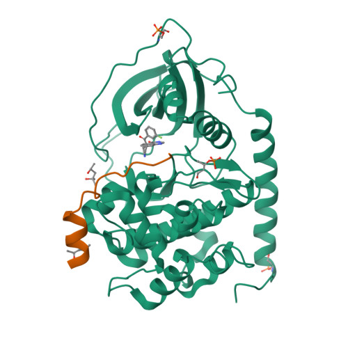
Protein Kinase A in complex with an Inhibitor


Experimental Data Snapshot

  • Method: X-RAY DIFFRACTION
  • Resolution: 1.95 Å
  • R-Value Free: 0.190 
  • R-Value Work: 0.155 
  • R-Value Observed: 0.156 

Starting Model: experimental
View more details

wwPDB Validation   3D Report Full Report


Ligand Structure Quality Assessment 


This is version 2.2 of the entry. See complete history


Literature

Addressing the Glycine-Rich Loop of Protein Kinases by a Multi-Facetted Interaction Network: Inhibition of Pka and a Pkb Mimic.

Lauber, B.S.Hardegger, L.A.Asraful, A.K.Lund, B.A.Dumele, O.Harder, M.Kuhn, B.Engh, R.A.Diederich, F.

(2016) Chemistry 22: 211

  • DOI: https://doi.org/10.1002/chem.201503552
  • Primary Citation of Related Structures:  
    4UJ1, 4UJ2, 4UJ9, 4UJA, 4UJB, 4Z83, 4Z84

  • PubMed Abstract: 

    Protein kinases continue to be hot targets in drug discovery research, as they are involved in many essential cellular processes and their deregulation can lead to a variety of diseases. A series of 32 enantiomerically pure inhibitors was synthesized and tested towards protein kinase A (PKA) and protein kinase B mimic PKAB3 (PKA triple mutant). The ligands bind to the hinge region, ribose pocket, and glycine-rich loop at the ATP site. Biological assays showed high potency against PKA, with Ki values in the low nanomolar range. The investigation demonstrates the significance of targeting the often neglected glycine-rich loop for gaining high binding potency. X-ray co-crystal structures revealed a multi-facetted network of ligand-loop interactions for the tightest binders, involving orthogonal dipolar contacts, sulfur and other dispersive contacts, amide-π stacking, and H-bonding to organofluorine, besides efficient water replacement. The network was analyzed in a computational approach.


  • Organizational Affiliation

    Laboratorium für Organische Chemie, ETH Zürich, Vladimir-Prelog-Weg 3, 8093 Zürich (Switzerland), Fax: (+41) 44-632-11-09.


Macromolecules
Find similar proteins by:  (by identity cutoff)  |  3D Structure
Entity ID: 1
MoleculeChains Sequence LengthOrganismDetailsImage
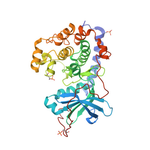
CAMP-DEPENDENT PROTEIN KINASE CATALYTIC SUBUNIT ALPHA351Homo sapiensMutation(s): 0 
EC: 2.7.11.11
UniProt & NIH Common Fund Data Resources
Find proteins for P17612 (Homo sapiens)
Explore P17612 
Go to UniProtKB:  P17612
PHAROS:  P17612
GTEx:  ENSG00000072062 
Entity Groups  
Sequence Clusters30% Identity50% Identity70% Identity90% Identity95% Identity100% Identity
UniProt GroupP17612
Sequence Annotations
Expand
  • Reference Sequence

Find similar proteins by:  Sequence   |   3D Structure  

Entity ID: 2
MoleculeChains Sequence LengthOrganismDetailsImage
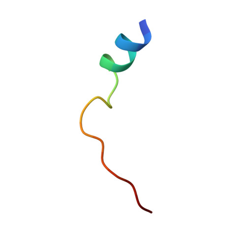
PEPTIDE20Homo sapiensMutation(s): 0 
UniProt & NIH Common Fund Data Resources
Find proteins for P61925 (Homo sapiens)
Explore P61925 
Go to UniProtKB:  P61925
PHAROS:  P61925
GTEx:  ENSG00000171033 
Entity Groups  
Sequence Clusters30% Identity50% Identity70% Identity90% Identity95% Identity100% Identity
UniProt GroupP61925
Sequence Annotations
Expand
  • Reference Sequence
Small Molecules
Ligands 2 Unique
IDChains Name / Formula / InChI Key2D Diagram3D Interactions
8BQ
Query on 8BQ

Download Ideal Coordinates CCD File 
C [auth A]7-[(3S,4R)-4-(3-fluorophenyl)carbonylpyrrolidin-3-yl]-3H-quinazolin-4-one
C19 H16 F N3 O2
POKNNHZYSUAARX-CVEARBPZSA-N
MPD
Query on MPD

Download Ideal Coordinates CCD File 
D [auth A],
E [auth B]
(4S)-2-METHYL-2,4-PENTANEDIOL
C6 H14 O2
SVTBMSDMJJWYQN-YFKPBYRVSA-N
Modified Residues  2 Unique
IDChains TypeFormula2D DiagramParent
SEP
Query on SEP
A
L-PEPTIDE LINKINGC3 H8 N O6 PSER
TPO
Query on TPO
A
L-PEPTIDE LINKINGC4 H10 N O6 PTHR
Experimental Data & Validation

Experimental Data

  • Method: X-RAY DIFFRACTION
  • Resolution: 1.95 Å
  • R-Value Free: 0.190 
  • R-Value Work: 0.155 
  • R-Value Observed: 0.156 
  • Space Group: P 21 21 21
Unit Cell:
Length ( Å )Angle ( ˚ )
a = 72.62α = 90
b = 75.371β = 90
c = 80.349γ = 90
Software Package:
Software NamePurpose
PHENIXrefinement
XDSdata reduction
XDSdata scaling
PHENIXphasing

Structure Validation

View Full Validation Report


Ligand Structure Quality Assessment 


Entry History 

Deposition Data

Revision History  (Full details and data files)

  • Version 1.0: 2016-04-13
    Type: Initial release
  • Version 2.0: 2019-01-30
    Changes: Atomic model, Data collection, Experimental preparation
  • Version 2.1: 2019-02-06
    Changes: Data collection, Experimental preparation
  • Version 2.2: 2024-01-10
    Changes: Data collection, Database references, Derived calculations, Other, Refinement description