4UCG | pdb_00004ucg

NmeDAH7PS R126S variant


Experimental Data Snapshot

  • Method: X-RAY DIFFRACTION
  • Resolution: 2.00 Å
  • R-Value Free: 
    0.220 (Depositor), 0.227 (DCC) 
  • R-Value Work: 
    0.180 (Depositor), 0.190 (DCC) 
  • R-Value Observed: 
    0.182 (Depositor) 

Starting Model: experimental
View more details

wwPDB Validation 3D Report Full Report

Validation slider image for 4UCG

Ligand Structure Quality Assessment 


This is version 1.2 of the entry. See complete history

Literature

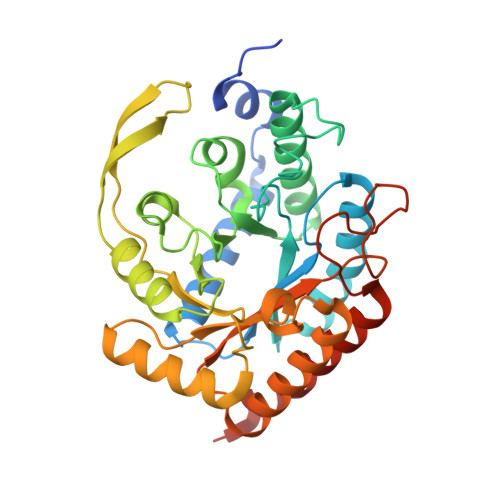
The Functional Unit of Neisseria Meningitidis 3-Deoxy-D-Arabino-Heptulosonate 7-Phosphate Synthase is Dimeric.

Cross, P.J.Heyes, L.C.Zhang, S.Nazmi, A.R.

(2016) PLoS One 11: 45187

  • DOI: https://doi.org/10.1371/journal.pone.0145187
  • Primary Citation Related Structures: 
    4UCG, 5DCE

  • PubMed Abstract: 

    Neisseria meningitidis 3-deoxy-D-arabino-heptulosonate 7-phosphate synthase (NmeDAH7PS) adopts a homotetrameric structure consisting of an extensive and a less extensive interface. Perturbation of the less extensive interface through a single mutation of a salt bridge (Arg126-Glu27) formed at the tetramer interface of all chains resulted in a dimeric DAH7PS in solution, as determined by small angle X-ray scattering, analytical ultracentrifugation and analytical size-exclusion chromatography. The dimeric NmeDAH7PSR126S variant was shown to be catalytically active in the aldol-like condensation reaction between D-erythrose 4-phosphate and phosphoenolpyruvate, and allosterically inhibited by L-phenylalanine to the same extent as the wild-type enzyme. The dimeric NmeDAH7PSR126S variant exhibited a slight reduction in thermal stability by differential scanning calorimetry experiments and a slow loss of activity over time compared to the wild-type enzyme. Although NmeDAH7PSR126S crystallised as a tetramer, like the wild-type enzyme, structural asymmetry at the less extensive interface was observed consistent with its destabilisation. The tetrameric association enabled by this Arg126-Glu27 salt-bridge appears to contribute solely to the stability of the protein, ultimately revealing that the functional unit of NmeDAH7PS is dimeric.


  • Organizational Affiliation
    • Biomolecular Interaction Centre and Department of Chemistry, University of Canterbury, Christchurch, New Zealand.

Macromolecule Content 

  • Total Structure Weight: 156.17 kDa 
  • Atom Count: 11,040 
  • Modeled Residue Count: 1,339 
  • Deposited Residue Count: 1,404 
  • Unique protein chains: 1

Macromolecules

Find similar proteins by:|  3D Structure
Entity ID: 1
MoleculeChains  Sequence LengthOrganismDetailsImage
PHOSPHO-2-DEHYDRO-3-DEOXYHEPTONATE ALDOLASE
A, B, C, D
351Neisseria meningitidisMutation(s): 1 
EC: 2.5.1.54
UniProt
Find proteins for Q9K169 (Neisseria meningitidis serogroup B (strain ATCC BAA-335 / MC58))
Explore Q9K169 
Go to UniProtKB:  Q9K169
Entity Groups
Sequence Clusters30% Identity50% Identity70% Identity90% Identity95% Identity100% Identity
UniProt GroupQ9K169
Sequence Annotations
Expand
Reference Sequence

Small Molecules

Ligands 4 Unique
IDChains Name / Formula / InChI Key2D Diagram3D Interactions
PEP

Query on PEP


Download:Ideal Coordinates CCD File
F [auth A],
K [auth B],
P [auth C],
R [auth D]
PHOSPHOENOLPYRUVATE
C3 H5 O6 P
DTBNBXWJWCWCIK-UHFFFAOYSA-N
PEG

Query on PEG


Download:Ideal Coordinates CCD File
H [auth A],
I [auth A],
M [auth B],
N [auth B]
DI(HYDROXYETHYL)ETHER
C4 H10 O3
MTHSVFCYNBDYFN-UHFFFAOYSA-N
SO4

Query on SO4


Download:Ideal Coordinates CCD File
G [auth A],
L [auth B],
S [auth D]
SULFATE ION
O4 S
QAOWNCQODCNURD-UHFFFAOYSA-L
MN

Query on MN


Download:Ideal Coordinates CCD File
E [auth A],
J [auth B],
O [auth C],
Q [auth D]
MANGANESE (II) ION
Mn
WAEMQWOKJMHJLA-UHFFFAOYSA-N

Experimental Data & Validation

Experimental Data

  • Method: X-RAY DIFFRACTION
  • Resolution: 2.00 Å
  • R-Value Free:  0.220 (Depositor), 0.227 (DCC) 
  • R-Value Work:  0.180 (Depositor), 0.190 (DCC) 
  • R-Value Observed: 0.182 (Depositor) 
Space Group: P 21 21 21
Unit Cell:
Length ( Å )Angle ( ˚ )
a = 78.949α = 90
b = 132.66β = 90
c = 147.722γ = 90
Software Package:
Software NamePurpose
REFMACrefinement
MOSFLMdata reduction
SCALAdata scaling
PHASERphasing

Structure Validation

View Full Validation Report


Ligand Structure Quality Assessment 


Entry History 

Deposition Data

Revision History  (Full details and data files)

  • Version 1.0: 2016-01-13
    Type: Initial release
  • Version 1.1: 2016-02-10
    Changes: Database references
  • Version 1.2: 2023-12-20
    Changes: Data collection, Database references, Derived calculations, Other, Refinement description