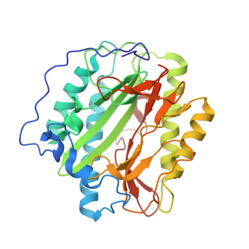
4U6Z

HsMetAP(F309M) in complex with 1-amino-2-propylpentyl)phosphonic acid


Experimental Data Snapshot

  • Method: X-RAY DIFFRACTION
  • Resolution: 1.80 Å
  • R-Value Free: 0.204 
  • R-Value Work: 0.167 
  • R-Value Observed: 0.169 

wwPDB Validation   3D Report Full Report


Ligand Structure Quality Assessment 


This is version 1.2 of the entry. See complete history


Literature

Identification of the Molecular Basis of Inhibitor Selectivity between the Human and Streptococcal Type I Methionine Aminopeptidases

Arya, T.Reddi, R.Kishor, C.Ganji, R.J.Bhukya, S.Gumpena, R.McGowan, S.Drag, M.Addlagatta, A.

(2015) J Med Chem 58: 2350-2357

  • DOI: https://doi.org/10.1021/jm501790e
  • Primary Citation of Related Structures:  
    4U1B, 4U69, 4U6C, 4U6E, 4U6J, 4U6W, 4U6Z, 4U70, 4U71, 4U73, 4U75, 4U76

  • PubMed Abstract: 

    The methionine aminopeptidase (MetAP) family is responsible for the cleavage of the initiator methionine from newly synthesized proteins. Currently, there are no small molecule inhibitors that show selectivity toward the bacterial MetAPs compared to the human enzyme. In our current study, we have screened 20 α-aminophosphonate derivatives and identified a molecule (compound 15) that selectively inhibits the S. pneumonia MetAP in low micromolar range but not the human enzyme. Further bioinformatics, biochemical, and structural analyses suggested that phenylalanine (F309) in the human enzyme and methionine (M205) in the S. pneumonia MetAP at the analogous position render them with different susceptibilities against the identified inhibitor. X-ray crystal structures of various inhibitors in complex with wild type and F309M enzyme further established the molecular basis for the inhibitor selectivity.


  • Organizational Affiliation

    Centre for Chemical Biology, CSIR-Indian Institute of Chemical Technology , Hyderabad 500 007, India.


Macromolecules
Find similar proteins by:  (by identity cutoff)  |  3D Structure
Entity ID: 1
MoleculeChains Sequence LengthOrganismDetailsImage
Methionine aminopeptidase 1306Homo sapiensMutation(s): 1 
Gene Names: METAP1KIAA0094
EC: 3.4.11.18
UniProt & NIH Common Fund Data Resources
Find proteins for P53582 (Homo sapiens)
Explore P53582 
Go to UniProtKB:  P53582
PHAROS:  P53582
GTEx:  ENSG00000164024 
Entity Groups  
Sequence Clusters30% Identity50% Identity70% Identity90% Identity95% Identity100% Identity
UniProt GroupP53582
Sequence Annotations
Expand
  • Reference Sequence
Small Molecules
Ligands 4 Unique
IDChains Name / Formula / InChI Key2D Diagram3D Interactions
Q07
Query on Q07

Download Ideal Coordinates CCD File 
F [auth A][(1R,2S)-1-amino-2-methylpentyl]phosphonic acid
C6 H16 N O3 P
AMCULSZUZIHVRH-NTSWFWBYSA-N
GOL
Query on GOL

Download Ideal Coordinates CCD File 
E [auth A]GLYCEROL
C3 H8 O3
PEDCQBHIVMGVHV-UHFFFAOYSA-N
CO
Query on CO

Download Ideal Coordinates CCD File 
B [auth A],
C [auth A]
COBALT (II) ION
Co
XLJKHNWPARRRJB-UHFFFAOYSA-N
K
Query on K

Download Ideal Coordinates CCD File 
D [auth A]POTASSIUM ION
K
NPYPAHLBTDXSSS-UHFFFAOYSA-N
Experimental Data & Validation

Experimental Data

  • Method: X-RAY DIFFRACTION
  • Resolution: 1.80 Å
  • R-Value Free: 0.204 
  • R-Value Work: 0.167 
  • R-Value Observed: 0.169 
  • Space Group: P 1 21 1
Unit Cell:
Length ( Å )Angle ( ˚ )
a = 47.137α = 90
b = 77.331β = 93.08
c = 47.153γ = 90
Software Package:
Software NamePurpose
REFMACrefinement

Structure Validation

View Full Validation Report


Ligand Structure Quality Assessment 


Entry History 

Deposition Data

Revision History  (Full details and data files)

  • Version 1.0: 2015-04-15
    Type: Initial release
  • Version 1.1: 2019-11-20
    Changes: Derived calculations, Source and taxonomy
  • Version 1.2: 2024-03-20
    Changes: Data collection, Database references