4U2H | pdb_00004u2h

The crystal structure of apo CalE6, a methionine gamma lyase from Micromonospora echinospora


Experimental Data Snapshot

  • Method: X-RAY DIFFRACTION
  • Resolution: 1.85 Å
  • R-Value Free: 
    0.232 (Depositor), 0.230 (DCC) 
  • R-Value Work: 
    0.209 (Depositor), 0.210 (DCC) 

wwPDB Validation   3D Report Full Report


This is version 1.1 of the entry. See complete history


Literature

Identification and Characterization of a Methionine gamma-Lyase in the Calicheamicin Biosynthetic Cluster of Micromonospora echinospora

Song, H.Xu, R.Guo, Z.

(2015) Chembiochem 16: 100-109

  • DOI: https://doi.org/10.1002/cbic.201402489
  • Primary Citation of Related Structures:  
    4U1T, 4U2H

  • PubMed Abstract: 

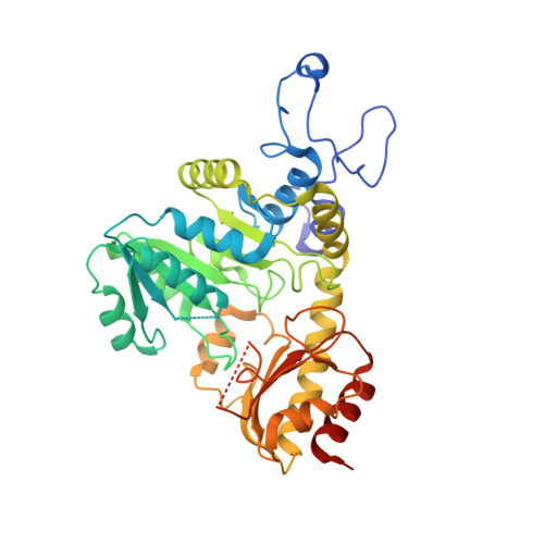
    CalE6 is a previously uncharacterized protein involved in the biosynthesis of calicheamicins in Micromonospora echinospora. It is a pyridoxal-5'-phosphate-dependent enzyme and exhibits high sequence homology to cystathionine γ-lyases and cystathionine γ-synthases. However, it was found to be active towards methionine and to convert this amino acid into α-ketobutyrate, ammonium, and methanethiol. The crystal structure of the cofactor-bound holoenzyme was resolved at 2.0 Å; it contains two active site residues, Gly105 and Val322, specific for methionine γ-lyases. Modeling of methionine into the active site allows identification of the active site residues responsible for substrate recognition and catalysis. These findings support that CalE6 is a putative methionine γ-lyase producing methanethiol as a building block in biosynthesis of calicheamicins.


  • Organizational Affiliation
    • Department of Chemistry and State Key Laboratory of Molecular Neuroscience, The Hong Kong University of Science and Technology, Clear Water Bay, Kowloon (Hong Kong).

Macromolecules
Find similar proteins by:  (by identity cutoff)  |  3D Structure
Entity ID: 1
MoleculeChains Sequence LengthOrganismDetailsImage
CalE6
A, B, C, D, E
A, B, C, D, E, F, G, H
389Micromonospora echinosporaMutation(s): 0 
Gene Names: calE6
EC: 4.4.1.11 (PDB Primary Data), 4.4.1.2 (UniProt)
UniProt
Find proteins for Q8KNG3 (Micromonospora echinospora)
Explore Q8KNG3 
Go to UniProtKB:  Q8KNG3
Entity Groups  
Sequence Clusters30% Identity50% Identity70% Identity90% Identity95% Identity100% Identity
UniProt GroupQ8KNG3
Sequence Annotations
Expand
  • Reference Sequence
Small Molecules
Ligands 1 Unique
IDChains Name / Formula / InChI Key2D Diagram3D Interactions
SO4
Query on SO4

Download Ideal Coordinates CCD File 
I [auth A]
J [auth A]
K [auth B]
L [auth B]
M [auth C]
I [auth A],
J [auth A],
K [auth B],
L [auth B],
M [auth C],
N [auth C],
O [auth D],
P [auth D],
Q [auth E],
R [auth E],
S [auth F],
T [auth F],
U [auth G],
V [auth G],
W [auth G],
X [auth H]
SULFATE ION
O4 S
QAOWNCQODCNURD-UHFFFAOYSA-L
Experimental Data & Validation

Experimental Data

  • Method: X-RAY DIFFRACTION
  • Resolution: 1.85 Å
  • R-Value Free:  0.232 (Depositor), 0.230 (DCC) 
  • R-Value Work:  0.209 (Depositor), 0.210 (DCC) 
Space Group: I 2 2 2
Unit Cell:
Length ( Å )Angle ( ˚ )
a = 145.204α = 90
b = 145.395β = 90
c = 346.119γ = 90
Software Package:
Software NamePurpose
HKL-2000data collection
HKL-2000data processing
HKL-2000data scaling
PHENIXrefinement
PHASERphasing
Cootmodel building

Structure Validation

View Full Validation Report


Entry History 

Deposition Data

Revision History  (Full details and data files)

  • Version 1.0: 2015-01-07
    Type: Initial release
  • Version 1.1: 2024-03-20
    Changes: Advisory, Data collection, Database references, Derived calculations, Other, Refinement description, Source and taxonomy