


Structural insights into modulation of MICAL activity by its Calponin Homology (CH) domain
Alqassim, S.S., Amzel, L.M., Bianchet, M.A.To be published.
Experimental Data Snapshot
Entity ID: 1 | |||||
|---|---|---|---|---|---|
| Molecule | Chains | Sequence Length | Organism | Details | Image |
| Protein-methionine sulfoxide oxidase MICAL1 | 615 | Mus musculus | Mutation(s): 0 Gene Names: Mical1, Mical, Nical EC: 1.14.13 (PDB Primary Data), 1.6.3.1 (UniProt), 1.14.13.225 (UniProt) | 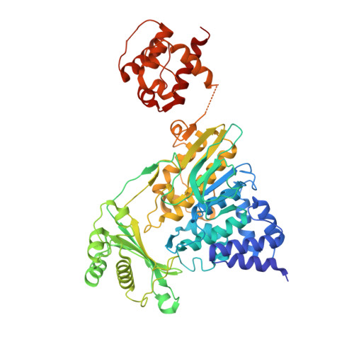 | |
UniProt | |||||
Entity Groups | |||||
| Sequence Clusters | 30% Identity50% Identity70% Identity90% Identity95% Identity100% Identity | ||||
| UniProt Group | Q8VDP3 | ||||
Sequence AnnotationsExpand | |||||
Reference Sequence | |||||
| Ligands 2 Unique | |||||
|---|---|---|---|---|---|
| ID | Chains | Name / Formula / InChI Key | 2D Diagram | 3D Interactions | |
| FAD Download:Ideal Coordinates CCD File | C [auth A] | FLAVIN-ADENINE DINUCLEOTIDE C27 H33 N9 O15 P2 VWWQXMAJTJZDQX-UYBVJOGSSA-N |  | ||
| PEG Download:Ideal Coordinates CCD File | B [auth A] | DI(HYDROXYETHYL)ETHER C4 H10 O3 MTHSVFCYNBDYFN-UHFFFAOYSA-N |  | ||
| Length ( Å ) | Angle ( ˚ ) |
|---|---|
| a = 70.233 | α = 90 |
| b = 50.174 | β = 101.16 |
| c = 96.966 | γ = 90 |
| Software Name | Purpose |
|---|---|
| PHENIX | refinement |
| HKL-2000 | data reduction |
| MOLREP | phasing |
| Coot | model building |
| PDB_EXTRACT | data extraction |
| HKL-2000 | data scaling |


