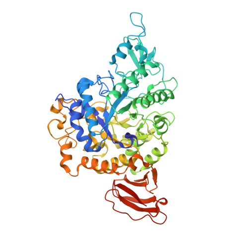
4TVU

Crystal structure of trehalose synthase from Deinococcus radiodurans reveals a closed conformation for catalysis of the intramolecular isomerization


Experimental Data Snapshot

  • Method: X-RAY DIFFRACTION
  • Resolution: 2.70 Å
  • R-Value Free: 0.236 
  • R-Value Work: 0.194 
  • R-Value Observed: 0.196 

Starting Model: experimental
View more details

wwPDB Validation   3D Report Full Report


This is version 1.1 of the entry. See complete history


Literature

Structures of trehalose synthase from Deinococcus radiodurans reveal that a closed conformation is involved in catalysis of the intramolecular isomerization.

Wang, Y.L.Chow, S.Y.Lin, Y.T.Hsieh, Y.C.Lee, G.C.Liaw, S.H.

(2014) Acta Crystallogr D Biol Crystallogr 70: 3144-3154

  • DOI: https://doi.org/10.1107/S1399004714022500
  • Primary Citation of Related Structures:  
    4TVU, 4WF7

  • PubMed Abstract: 

    Trehalose synthase catalyzes the simple conversion of the inexpensive maltose into trehalose with a side reaction of hydrolysis. Here, the crystal structures of the wild type and the N253A mutant of Deinococcus radiodurans trehalose synthase (DrTS) in complex with the inhibitor Tris are reported. DrTS consists of a catalytic (β/α)8 barrel, subdomain B, a C-terminal β domain and two TS-unique subdomains (S7 and S8). The C-terminal domain and S8 contribute the majority of the dimeric interface. DrTS shares high structural homology with sucrose hydrolase, amylosucrase and sucrose isomerase in complex with sucrose, in particular a virtually identical active-site architecture and a similar substrate-induced rotation of subdomain B. The inhibitor Tris was bound and mimics a sugar at the -1 subsite. A maltose was modelled into the active site, and subsequent mutational analysis suggested that Tyr213, Glu320 and Glu324 are essential within the +1 subsite for the TS activity. In addition, the interaction networks between subdomains B and S7 seal the active-site entrance. Disruption of such networks through the replacement of Arg148 and Asn253 with alanine resulted in a decrease in isomerase activity by 8-9-fold and an increased hydrolase activity by 1.5-1.8-fold. The N253A structure showed a small pore created for water entry. Therefore, our DrTS-Tris may represent a substrate-induced closed conformation that will facilitate intramolecular isomerization and minimize disaccharide hydrolysis.


  • Organizational Affiliation

    Institute of Biochemistry and Molecular Biology, National Yang-Ming University, Taipei 11221, Taiwan.


Macromolecules
Find similar proteins by:  (by identity cutoff)  |  3D Structure
Entity ID: 1
MoleculeChains Sequence LengthOrganismDetailsImage
Trehalose synthase
A, B, C, D, E
A, B, C, D, E, F, G, H
571Deinococcus radiodurans R1 = ATCC 13939 = DSM 20539Mutation(s): 3 
EC: 5.4.99.16
UniProt
Find proteins for I3NX86 (Deinococcus radiodurans (strain ATCC 13939 / DSM 20539 / JCM 16871 / CCUG 27074 / LMG 4051 / NBRC 15346 / NCIMB 9279 / VKM B-1422 / R1))
Explore I3NX86 
Go to UniProtKB:  I3NX86
Entity Groups  
Sequence Clusters30% Identity50% Identity70% Identity90% Identity95% Identity100% Identity
UniProt GroupI3NX86
Sequence Annotations
Expand
  • Reference Sequence
Small Molecules
Ligands 3 Unique
IDChains Name / Formula / InChI Key2D Diagram3D Interactions
TRS
Query on TRS

Download Ideal Coordinates CCD File 
CA [auth G]
FA [auth H]
K [auth A]
N [auth B]
Q [auth C]
CA [auth G],
FA [auth H],
K [auth A],
N [auth B],
Q [auth C],
T [auth D],
W [auth E],
Z [auth F]
2-AMINO-2-HYDROXYMETHYL-PROPANE-1,3-DIOL
C4 H12 N O3
LENZDBCJOHFCAS-UHFFFAOYSA-O
CA
Query on CA

Download Ideal Coordinates CCD File 
AA [auth G]
DA [auth H]
I [auth A]
L [auth B]
O [auth C]
AA [auth G],
DA [auth H],
I [auth A],
L [auth B],
O [auth C],
R [auth D],
U [auth E],
X [auth F]
CALCIUM ION
Ca
BHPQYMZQTOCNFJ-UHFFFAOYSA-N
MG
Query on MG

Download Ideal Coordinates CCD File 
BA [auth G]
EA [auth H]
J [auth A]
M [auth B]
P [auth C]
BA [auth G],
EA [auth H],
J [auth A],
M [auth B],
P [auth C],
S [auth D],
V [auth E],
Y [auth F]
MAGNESIUM ION
Mg
JLVVSXFLKOJNIY-UHFFFAOYSA-N
Experimental Data & Validation

Experimental Data

  • Method: X-RAY DIFFRACTION
  • Resolution: 2.70 Å
  • R-Value Free: 0.236 
  • R-Value Work: 0.194 
  • R-Value Observed: 0.196 
  • Space Group: P 1 21 1
Unit Cell:
Length ( Å )Angle ( ˚ )
a = 95.609α = 90
b = 195.896β = 92.89
c = 130.986γ = 90
Software Package:
Software NamePurpose
REFMACrefinement
SCALEPACKdata reduction
PDB_EXTRACTdata extraction
SCALEPACKdata scaling

Structure Validation

View Full Validation Report


Entry History & Funding Information

Deposition Data


Funding OrganizationLocationGrant Number
National Science CouncilTaiwanNSC 102-2311-B-101-005-

Revision History  (Full details and data files)

  • Version 1.0: 2014-12-17
    Type: Initial release
  • Version 1.1: 2023-11-08
    Changes: Data collection, Database references, Derived calculations, Other, Refinement description, Source and taxonomy