4TVP | pdb_00004tvp

Crystal Structure of the HIV-1 BG505 SOSIP.664 Env Trimer Ectodomain, Comprising Atomic-Level Definition of Pre-Fusion gp120 and gp41, in Complex with Human Antibodies PGT122 and 35O22


Experimental Data Snapshot

  • Method: X-RAY DIFFRACTION
  • Resolution: 3.10 Å
  • R-Value Free: 
    0.248 (Depositor), 0.247 (DCC) 
  • R-Value Work: 
    0.213 (Depositor), 0.217 (DCC) 
  • R-Value Observed: 
    0.215 (Depositor) 

Starting Models: experimental
View more details

wwPDB Validation 3D Report Full Report

Validation slider image for 4TVP

Ligand Structure Quality Assessment 


This is version 3.2 of the entry. See complete history

Literature

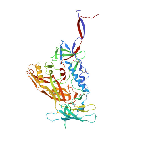
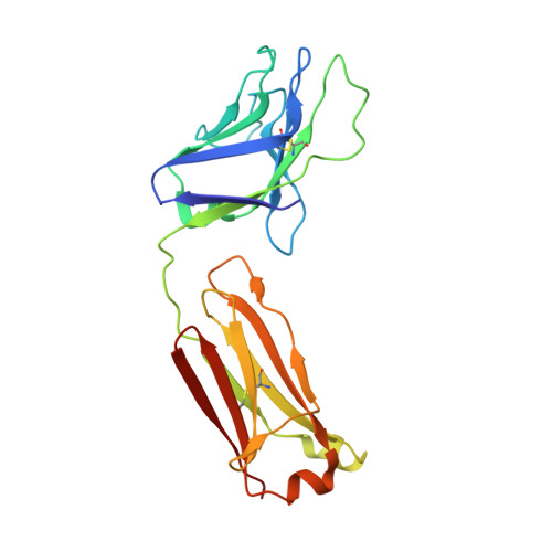
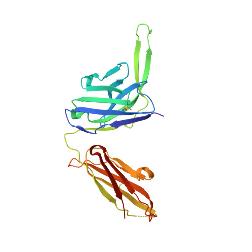
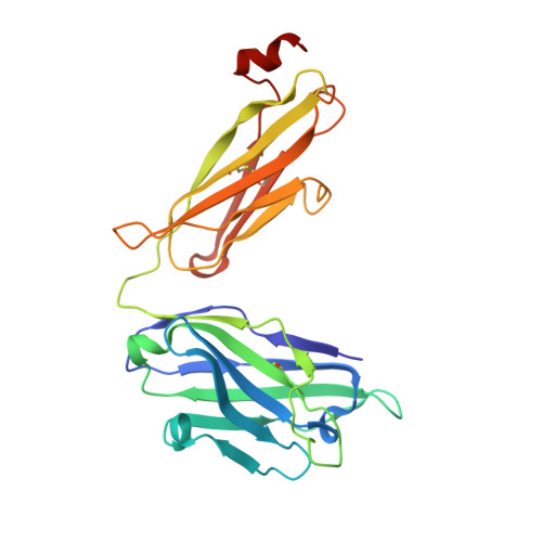
Structure and immune recognition of trimeric pre-fusion HIV-1 Env.

Pancera, M.Zhou, T.Druz, A.Georgiev, I.S.Soto, C.Gorman, J.Huang, J.Acharya, P.Chuang, G.Y.Ofek, G.Stewart-Jones, G.B.Stuckey, J.Bailer, R.T.Joyce, M.G.Louder, M.K.Tumba, N.Yang, Y.Zhang, B.Cohen, M.S.Haynes, B.F.Mascola, J.R.Morris, L.Munro, J.B.Blanchard, S.C.Mothes, W.Connors, M.Kwong, P.D.

(2014) Nature 514: 455-461

  • DOI: https://doi.org/10.1038/nature13808
  • Primary Citation Related Structures: 
    4TVP

  • PubMed Abstract: 

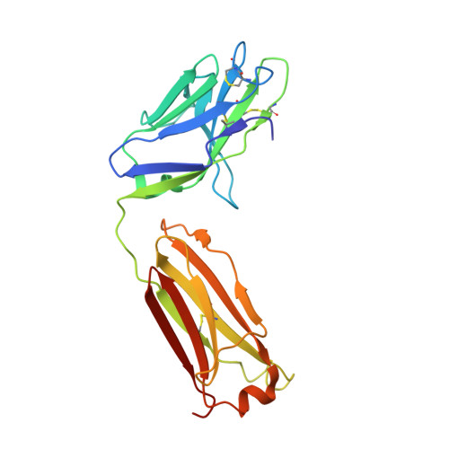
    The human immunodeficiency virus type 1 (HIV-1) envelope (Env) spike, comprising three gp120 and three gp41 subunits, is a conformational machine that facilitates HIV-1 entry by rearranging from a mature unliganded state, through receptor-bound intermediates, to a post-fusion state. As the sole viral antigen on the HIV-1 virion surface, Env is both the target of neutralizing antibodies and a focus of vaccine efforts. Here we report the structure at 3.5 Å resolution for an HIV-1 Env trimer captured in a mature closed state by antibodies PGT122 and 35O22. This structure reveals the pre-fusion conformation of gp41, indicates rearrangements needed for fusion activation, and defines parameters of immune evasion and immune recognition. Pre-fusion gp41 encircles amino- and carboxy-terminal strands of gp120 with four helices that form a membrane-proximal collar, fastened by insertion of a fusion peptide-proximal methionine into a gp41-tryptophan clasp. Spike rearrangements required for entry involve opening the clasp and expelling the termini. N-linked glycosylation and sequence-variable regions cover the pre-fusion closed spike; we used chronic cohorts to map the prevalence and location of effective HIV-1-neutralizing responses, which were distinguished by their recognition of N-linked glycan and tolerance for epitope-sequence variation.


  • Organizational Affiliation
    • Vaccine Research Center, National Institute of Allergy and Infectious Diseases, National Institutes of Health, Bethesda, Maryland 20892, USA.

Macromolecule Content 

  • Total Structure Weight: 181.42 kDa 
  • Atom Count: 12,188 
  • Modeled Residue Count: 1,472 
  • Deposited Residue Count: 1,541 
  • Unique protein chains: 6

Macromolecules

Find similar proteins by:|  3D Structure
Entity ID: 1
MoleculeChains  Sequence LengthOrganismDetailsImage
Envelope glycoprotein gp160A [auth G]481Human immunodeficiency virus 1Mutation(s): 2 
Gene Names: env
UniProt
Find proteins for Q2N0S5 (Human immunodeficiency virus type 1)
Explore Q2N0S5 
Go to UniProtKB:  Q2N0S5
Entity Groups
Sequence Clusters30% Identity50% Identity70% Identity90% Identity95% Identity100% Identity
UniProt GroupQ2N0S5
Glycosylation
Glycosylation Sites: 18
Sequence Annotations
Expand
Reference Sequence
Find similar proteins by:|  3D Structure
Entity ID: 2
MoleculeChains  Sequence LengthOrganismDetailsImage
Envelope glycoprotein gp160153Human immunodeficiency virus 1Mutation(s): 2 
Gene Names: env
UniProt
Find proteins for Q2N0S5 (Human immunodeficiency virus type 1)
Explore Q2N0S5 
Go to UniProtKB:  Q2N0S5
Entity Groups
Sequence Clusters30% Identity50% Identity70% Identity90% Identity95% Identity100% Identity
UniProt GroupQ2N0S5
Glycosylation
Glycosylation Sites: 3
Sequence Annotations
Expand
Reference Sequence
Find similar proteins by:|  3D Structure
Entity ID: 3
MoleculeChains  Sequence LengthOrganismDetailsImage
PGT122 Light chainC [auth L]213Homo sapiensMutation(s): 0 
Find similar proteins by:|  3D Structure
Entity ID: 4
MoleculeChains  Sequence LengthOrganismDetailsImage
PGT122 Heavy chainD [auth H]235Homo sapiensMutation(s): 0 
Find similar proteins by:|  3D Structure
Entity ID: 5
MoleculeChains  Sequence LengthOrganismDetailsImage
35O22 Heavy chainE [auth D]243Homo sapiensMutation(s): 0 
Find similar proteins by:|  3D Structure
Entity ID: 6
MoleculeChains  Sequence LengthOrganismDetailsImage
35O22 Light chainF [auth E]216Homo sapiensMutation(s): 0 

Oligosaccharides

Help  
Entity ID: 7
MoleculeChains Length2D Diagram GlycosylationD Interactions
alpha-D-mannopyranose-(1-3)-[alpha-D-mannopyranose-(1-6)]alpha-D-mannopyranose-(1-6)-[alpha-D-mannopyranose-(1-3)]beta-D-mannopyranose-(1-4)-2-acetamido-2-deoxy-beta-D-glucopyranose-(1-4)-2-acetamido-2-deoxy-beta-D-glucopyranoseG [auth A]7N-Glycosylation
Glycosylation Resources
GlyTouCan: G55220VL
GlyCosmos: G55220VL
GlyGen: G55220VL
Entity ID: 8
MoleculeChains Length2D Diagram GlycosylationD Interactions
alpha-D-mannopyranose-(1-3)-beta-D-mannopyranose-(1-4)-2-acetamido-2-deoxy-beta-D-glucopyranose-(1-4)-2-acetamido-2-deoxy-beta-D-glucopyranoseH [auth C]4N-Glycosylation
Glycosylation Resources
GlyTouCan: G81315DD
GlyCosmos: G81315DD
GlyGen: G81315DD
Entity ID: 9
MoleculeChains Length2D Diagram GlycosylationD Interactions
2-acetamido-2-deoxy-beta-D-glucopyranose-(1-4)-2-acetamido-2-deoxy-beta-D-glucopyranose
I [auth F],
K [auth J],
L [auth K],
M,
P,
I [auth F],
K [auth J],
L [auth K],
M,
P,
Q,
R,
S
2N-Glycosylation
Glycosylation Resources
GlyTouCan: G42666HT
GlyCosmos: G42666HT
GlyGen: G42666HT
Entity ID: 10
MoleculeChains Length2D Diagram GlycosylationD Interactions
alpha-D-mannopyranose-(1-2)-alpha-D-mannopyranose-(1-3)-[alpha-D-mannopyranose-(1-6)]beta-D-mannopyranose-(1-4)-2-acetamido-2-deoxy-beta-D-glucopyranose-(1-4)-2-acetamido-2-deoxy-beta-D-glucopyranoseJ [auth I]6N-Glycosylation
Glycosylation Resources
GlyTouCan: G56014GC
GlyCosmos: G56014GC
GlyGen: G56014GC
Entity ID: 11
MoleculeChains Length2D Diagram GlycosylationD Interactions
beta-D-mannopyranose-(1-4)-2-acetamido-2-deoxy-beta-D-glucopyranose-(1-4)-2-acetamido-2-deoxy-beta-D-glucopyranose
N
3N-Glycosylation
Glycosylation Resources
GlyTouCan: G15407YE
GlyCosmos: G15407YE
GlyGen: G15407YE
Entity ID: 12
MoleculeChains Length2D Diagram GlycosylationD Interactions
alpha-D-mannopyranose-(1-3)-[alpha-D-mannopyranose-(1-6)]beta-D-mannopyranose-(1-4)-2-acetamido-2-deoxy-beta-D-glucopyranose-(1-4)-2-acetamido-2-deoxy-beta-D-glucopyranose
O
5N-Glycosylation
Glycosylation Resources
GlyTouCan: G22768VO
GlyCosmos: G22768VO
GlyGen: G22768VO
Entity ID: 13
MoleculeChains Length2D Diagram GlycosylationD Interactions
alpha-D-mannopyranose-(1-2)-alpha-D-mannopyranose-(1-2)-alpha-D-mannopyranose-(1-3)-[alpha-D-mannopyranose-(1-2)-alpha-D-mannopyranose-(1-3)-[alpha-D-mannopyranose-(1-6)]alpha-D-mannopyranose-(1-6)]beta-D-mannopyranose-(1-4)-2-acetamido-2-deoxy-beta-D-glucopyranose-(1-4)-2-acetamido-2-deoxy-beta-D-glucopyranose
T
10N-Glycosylation
Glycosylation Resources
GlyTouCan: G91704UR
GlyCosmos: G91704UR
GlyGen: G91704UR

Small Molecules

Ligands 2 Unique
IDChains Name / Formula / InChI Key2D Diagram3D Interactions
NAG

Query on NAG


Download:Ideal Coordinates CCD File
FA [auth B]
GA [auth B]
HA [auth B]
LA [auth H]
U [auth G]
FA [auth B],
GA [auth B],
HA [auth B],
LA [auth H],
U [auth G],
V [auth G],
W [auth G],
X [auth G]
2-acetamido-2-deoxy-beta-D-glucopyranose
C8 H15 N O6
OVRNDRQMDRJTHS-FMDGEEDCSA-N
SO4

Query on SO4


Download:Ideal Coordinates CCD File
AA [auth G]
BA [auth G]
CA [auth G]
DA [auth G]
EA [auth G]
AA [auth G],
BA [auth G],
CA [auth G],
DA [auth G],
EA [auth G],
IA [auth B],
JA [auth B],
KA [auth L],
Y [auth G],
Z [auth G]
SULFATE ION
O4 S
QAOWNCQODCNURD-UHFFFAOYSA-L

Experimental Data & Validation

Experimental Data

  • Method: X-RAY DIFFRACTION
  • Resolution: 3.10 Å
  • R-Value Free:  0.248 (Depositor), 0.247 (DCC) 
  • R-Value Work:  0.213 (Depositor), 0.217 (DCC) 
  • R-Value Observed: 0.215 (Depositor) 
Space Group: P 63
Unit Cell:
Length ( Å )Angle ( ˚ )
a = 128.89α = 90
b = 128.89β = 90
c = 313.42γ = 120
Software Package:
Software NamePurpose
PHENIXrefinement
HKL-2000data scaling
PHASERphasing

Structure Validation

View Full Validation Report


Ligand Structure Quality Assessment 


Entry History 

Deposition Data

Revision History  (Full details and data files)

  • Version 1.0: 2014-10-08
    Type: Initial release
  • Version 1.1: 2014-10-22
    Changes: Database references
  • Version 1.2: 2014-11-05
    Changes: Database references
  • Version 1.3: 2015-04-22
    Changes: Structure summary
  • Version 2.0: 2017-11-22
    Changes: Atomic model, Database references, Derived calculations, Other, Refinement description, Source and taxonomy, Structure summary
  • Version 3.0: 2020-07-29
    Type: Remediation
    Reason: Carbohydrate remediation
    Changes: Atomic model, Data collection, Derived calculations, Refinement description, Structure summary
  • Version 3.1: 2023-09-27
    Changes: Data collection, Database references, Derived calculations, Refinement description, Structure summary
  • Version 3.2: 2024-10-30
    Changes: Structure summary