4TVC | pdb_00004tvc

N-terminally truncated dextransucrase DSR-E from Leuconostoc mesenteroides NRRL B-1299 in complex with gluco-oligosaccharides


Experimental Data Snapshot

  • Method: X-RAY DIFFRACTION
  • Resolution: 1.85 Å
  • R-Value Free: 
    0.205 (Depositor), 0.213 (DCC) 
  • R-Value Work: 
    0.161 (Depositor), 0.170 (DCC) 
  • R-Value Observed: 
    0.163 (Depositor) 

Starting Model: experimental
View more details

wwPDB Validation 3D Report Full Report

Validation slider image for 4TVC

Ligand Structure Quality Assessment 


This is version 2.2 of the entry. See complete history

Literature

Structural Insights into the Carbohydrate Binding Ability of an alpha-(12) Branching Sucrase from Glycoside Hydrolase Family 70.

Brison, Y.Malbert, Y.Czaplicki, G.Mourey, L.Remaud-Simeon, M.Tranier, S.

(2016) J Biological Chem 291: 7527-7540

  • DOI: https://doi.org/10.1074/jbc.M115.688796
  • Primary Citation Related Structures: 
    4TTU, 4TVC, 4TVD

  • PubMed Abstract: 

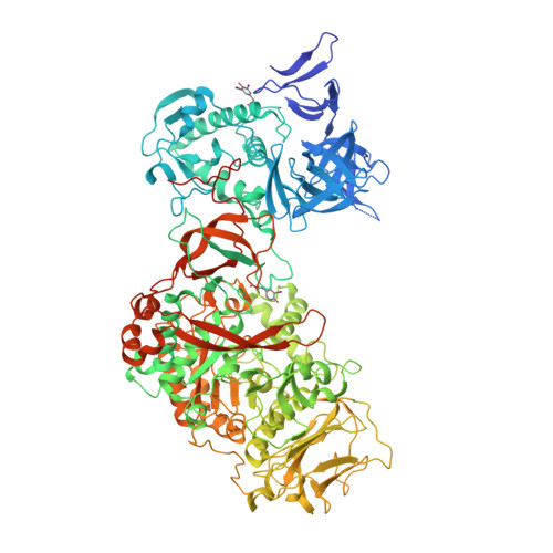
    The α-(1→2) branching sucrase ΔN123-GBD-CD2 is a transglucosylase belonging to glycoside hydrolase family 70 (GH70) that catalyzes the transfer ofd-glucosyl units from sucroseto dextrans or gluco-oligosaccharides via the formation of α-(1→2) glucosidic linkages. The first structures of ΔN123-GBD-CD2 in complex withd-glucose, isomaltosyl, or isomaltotriosyl residues were solved. The glucose complex revealed three glucose-binding sites in the catalytic gorge and six additional binding sites at the surface of domains B, IV, and V. Soaking with isomaltotriose or gluco-oligosaccharides led to structures in which isomaltosyl or isomaltotriosyl residues were found in glucan binding pockets located in domain V. One aromatic residue is systematically identified at the bottom of these pockets in stacking interaction with one glucosyl moiety. The carbohydrate is also maintained by a network of hydrogen bonds and van der Waals interactions. The sequence of these binding pockets is conserved and repeatedly present in domain V of several GH70 glucansucrases known to bind α-glucans. These findings provide the first structural evidence of the molecular interaction occurring between isomalto-oligosaccharides and domain V of the GH70 enzymes.


  • Organizational Affiliation
    • the INRA, UMR792 Ingénierie des Systèmes Biologiques et des Procédés, F-31400 Toulouse.

Macromolecule Content 

  • Total Structure Weight: 124.87 kDa 
  • Atom Count: 9,183 
  • Modeled Residue Count: 1,036 
  • Deposited Residue Count: 1,108 
  • Unique protein chains: 1

Macromolecules

Find similar proteins by:|  3D Structure
Entity ID: 1
MoleculeChains  Sequence LengthOrganismDetailsImage
Dextransucrase1,108Leuconostoc mesenteroidesMutation(s): 0 
Gene Names: dsrE
EC: 2.4.1.5
UniProt
Find proteins for Q8G9Q2 (Leuconostoc mesenteroides)
Explore Q8G9Q2 
Go to UniProtKB:  Q8G9Q2
Entity Groups
Sequence Clusters30% Identity50% Identity70% Identity90% Identity95% Identity100% Identity
UniProt GroupQ8G9Q2
Sequence Annotations
Expand
Reference Sequence

Oligosaccharides

Help  
Entity ID: 2
MoleculeChains Length2D Diagram GlycosylationD Interactions
alpha-D-glucopyranose-(1-6)-alpha-D-glucopyranose-(1-6)-alpha-D-glucopyranose
B
3N/A
Glycosylation Resources
GlyTouCan: G02998EX
GlyCosmos: G02998EX
GlyGen: G02998EX

Small Molecules

Ligands 4 Unique
IDChains Name / Formula / InChI Key2D Diagram3D Interactions
GLC

Query on GLC


Download:Ideal Coordinates CCD File
E [auth A]alpha-D-glucopyranose
C6 H12 O6
WQZGKKKJIJFFOK-DVKNGEFBSA-N
GOL

Query on GOL


Download:Ideal Coordinates CCD File
F [auth A],
G [auth A],
H [auth A]
GLYCEROL
C3 H8 O3
PEDCQBHIVMGVHV-UHFFFAOYSA-N
CA

Query on CA


Download:Ideal Coordinates CCD File
C [auth A]CALCIUM ION
Ca
BHPQYMZQTOCNFJ-UHFFFAOYSA-N
NA

Query on NA


Download:Ideal Coordinates CCD File
D [auth A]SODIUM ION
Na
FKNQFGJONOIPTF-UHFFFAOYSA-N
Modified Residues  2 Unique
IDChains TypeFormula2D DiagramParent
IYR
Query on IYR
A
L-PEPTIDE LINKINGC9 H10 I N O3TYR
TYI
Query on TYI
A
L-PEPTIDE LINKINGC9 H9 I2 N O3TYR

Experimental Data & Validation

Experimental Data

  • Method: X-RAY DIFFRACTION
  • Resolution: 1.85 Å
  • R-Value Free:  0.205 (Depositor), 0.213 (DCC) 
  • R-Value Work:  0.161 (Depositor), 0.170 (DCC) 
  • R-Value Observed: 0.163 (Depositor) 
Space Group: P 21 21 21
Unit Cell:
Length ( Å )Angle ( ˚ )
a = 68.31α = 90
b = 100.14β = 90
c = 186.24γ = 90
Software Package:
Software NamePurpose
REFMACrefinement
iMOSFLMdata reduction
SCALAdata scaling
PHASERphasing

Structure Validation

View Full Validation Report


Ligand Structure Quality Assessment 


Entry History 

Deposition Data

Revision History  (Full details and data files)

  • Version 1.0: 2015-07-29
    Type: Initial release
  • Version 1.1: 2016-02-24
    Changes: Database references
  • Version 1.2: 2016-04-13
    Changes: Database references
  • Version 2.0: 2020-07-29
    Type: Remediation
    Reason: Carbohydrate remediation
    Changes: Atomic model, Data collection, Derived calculations, Structure summary
  • Version 2.1: 2023-12-20
    Changes: Data collection, Database references, Derived calculations, Refinement description, Structure summary
  • Version 2.2: 2024-11-20
    Changes: Structure summary