4TTG | pdb_00004ttg

Beta-galactosidase (E. coli) in the presence of potassium chloride.


Experimental Data Snapshot

  • Method: X-RAY DIFFRACTION
  • Resolution: 1.60 Å
  • R-Value Free: 
    0.188 (Depositor), 0.193 (DCC) 
  • R-Value Work: 
    0.160 (Depositor) 
  • R-Value Observed: 
    0.160 (Depositor) 

Starting Model: experimental
View more details

wwPDB Validation 3D Report Full Report

Validation slider image for 4TTG

This is version 1.5 of the entry. See complete history

Literature

Elucidating factors important for monovalent cation selectivity in enzymes: E. coli beta-galactosidase as a model.

Wheatley, R.W.Juers, D.H.Lev, B.B.Huber, R.E.Noskov, S.Y.

(2015) Phys Chem Chem Phys 17: 10899-10909

  • DOI: https://doi.org/10.1039/c4cp04952g
  • Primary Citation Related Structures: 
    4TTG

  • PubMed Abstract: 

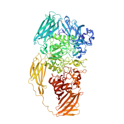
    Many enzymes require a specific monovalent cation (M(+)), that is either Na(+) or K(+), for optimal activity. While high selectivity M(+) sites in transport proteins have been extensively studied, enzyme M(+) binding sites generally have lower selectivity and are less characterized. Here we study the M(+) binding site of the model enzyme E. coli β-galactosidase, which is about 10 fold selective for Na(+) over K(+). Combining data from X-ray crystallography and computational models, we find the electrostatic environment predominates in defining the Na(+) selectivity. In this lower selectivity site rather subtle influences on the electrostatic environment become significant, including the induced polarization effects of the M(+) on the coordinating ligands and the effect of second coordination shell residues on the charge distribution of the primary ligands. This work expands the knowledge of ion selectivity in proteins to denote novel mechanisms important for the selectivity of M(+) sites in enzymes.


  • Organizational Affiliation
    • Division of Biochemistry, Department of Biological Sciences, University of Calgary, 2500 University Drive NW, Calgary, AB T2N 1N4, Canada.

Macromolecule Content 

  • Total Structure Weight: 477.47 kDa 
  • Atom Count: 38,364 
  • Modeled Residue Count: 4,060 
  • Deposited Residue Count: 4,092 
  • Unique protein chains: 1

Macromolecules

Find similar proteins by:|  3D Structure
Entity ID: 1
MoleculeChains  Sequence LengthOrganismDetailsImage
Beta-galactosidase
A, B, C, D
1,023Escherichia coliMutation(s): 0 
Gene Names: lacZ
EC: 3.2.1.23
UniProt
Find proteins for Q8VNN2 (Escherichia coli)
Explore Q8VNN2 
Go to UniProtKB:  Q8VNN2
Entity Groups
Sequence Clusters30% Identity50% Identity70% Identity90% Identity95% Identity100% Identity
UniProt GroupQ8VNN2
Sequence Annotations
Expand
Reference Sequence

Small Molecules

Ligands 4 Unique
IDChains Name / Formula / InChI Key2D Diagram3D Interactions
DMS

Query on DMS


Download:Ideal Coordinates CCD File
AA [auth A]
AC [auth B]
AD [auth C]
AE [auth C]
AF [auth D]
AA [auth A],
AC [auth B],
AD [auth C],
AE [auth C],
AF [auth D],
BA [auth A],
BC [auth B],
BD [auth C],
BE [auth C],
BF [auth D],
CA [auth A],
CC [auth B],
CD [auth C],
CE [auth C],
CF [auth D],
DA [auth A],
DB [auth B],
DC [auth B],
DD [auth C],
DE [auth C],
DF [auth D],
EA [auth A],
EB [auth B],
EC [auth B],
ED [auth C],
EE [auth D],
EF [auth D],
FA [auth A],
FB [auth B],
FC [auth B],
FD [auth C],
FF [auth D],
GA [auth A],
GB [auth B],
GC [auth B],
GD [auth C],
GF [auth D],
HA [auth A],
HB [auth B],
HC [auth B],
HD [auth C],
HF [auth D],
IA [auth A],
IB [auth B],
IC [auth B],
ID [auth C],
IF [auth D],
JA [auth A],
JB [auth B],
JC [auth B],
JD [auth C],
JF [auth D],
KA [auth A],
KB [auth B],
KC [auth B],
KD [auth C],
KF [auth D],
LA [auth A],
LB [auth B],
LD [auth C],
MA [auth A],
MB [auth B],
MD [auth C],
ME [auth D],
N [auth A],
NA [auth A],
NB [auth B],
ND [auth C],
NE [auth D],
O [auth A],
OA [auth A],
OB [auth B],
OD [auth C],
OE [auth D],
P [auth A],
PA [auth A],
PB [auth B],
PD [auth C],
PE [auth D],
Q [auth A],
QA [auth A],
QB [auth B],
QD [auth C],
QE [auth D],
R [auth A],
RA [auth A],
RB [auth B],
RD [auth C],
RE [auth D],
S [auth A],
SA [auth A],
SB [auth B],
SD [auth C],
SE [auth D],
T [auth A],
TA [auth B],
TB [auth B],
TC [auth C],
TD [auth C],
TE [auth D],
U [auth A],
UA [auth B],
UB [auth B],
UC [auth C],
UD [auth C],
UE [auth D],
V [auth A],
VA [auth B],
VB [auth B],
VC [auth C],
VD [auth C],
VE [auth D],
W [auth A],
WB [auth B],
WC [auth C],
WD [auth C],
WE [auth D],
X [auth A],
XB [auth B],
XC [auth C],
XD [auth C],
XE [auth D],
Y [auth A],
YB [auth B],
YC [auth C],
YD [auth C],
YE [auth D],
Z [auth A],
ZB [auth B],
ZC [auth C],
ZD [auth C],
ZE [auth D]
DIMETHYL SULFOXIDE
C2 H6 O S
IAZDPXIOMUYVGZ-UHFFFAOYSA-N
K

Query on K


Download:Ideal Coordinates CCD File
AB [auth B]
BB [auth B]
CB [auth B]
I [auth A]
IE [auth D]
AB [auth B],
BB [auth B],
CB [auth B],
I [auth A],
IE [auth D],
J [auth A],
JE [auth D],
K [auth A],
KE [auth D],
L [auth A],
LE [auth D],
PC [auth C],
QC [auth C],
RC [auth C],
SC [auth C],
ZA [auth B]
POTASSIUM ION
K
NPYPAHLBTDXSSS-UHFFFAOYSA-N
CL

Query on CL


Download:Ideal Coordinates CCD File
M [auth A]CHLORIDE ION
Cl
VEXZGXHMUGYJMC-UHFFFAOYSA-M
MG

Query on MG


Download:Ideal Coordinates CCD File
E [auth A]
F [auth A]
FE [auth D]
G [auth A]
GE [auth D]
E [auth A],
F [auth A],
FE [auth D],
G [auth A],
GE [auth D],
H [auth A],
HE [auth D],
LC [auth C],
MC [auth C],
NC [auth C],
OC [auth C],
WA [auth B],
XA [auth B],
YA [auth B]
MAGNESIUM ION
Mg
JLVVSXFLKOJNIY-UHFFFAOYSA-N
Modified Residues  1 Unique
IDChains TypeFormula2D DiagramParent
CSD
Query on CSD
A, B, C, D
L-PEPTIDE LINKINGC3 H7 N O4 SCYS

Experimental Data & Validation

Experimental Data

  • Method: X-RAY DIFFRACTION
  • Resolution: 1.60 Å
  • R-Value Free:  0.188 (Depositor), 0.193 (DCC) 
  • R-Value Work:  0.160 (Depositor) 
  • R-Value Observed: 0.160 (Depositor) 
Space Group: P 21 21 21
Unit Cell:
Length ( Å )Angle ( ˚ )
a = 149.29α = 90
b = 168.1β = 90
c = 200.31γ = 90
Software Package:
Software NamePurpose
REFMACrefinement

Structure Validation

View Full Validation Report


Entry History 

Deposition Data

  • Released Date: 2015-03-25 
  • Deposition Author(s): Juers, D.H.

Revision History  (Full details and data files)

  • Version 1.0: 2015-03-25
    Type: Initial release
  • Version 1.1: 2015-04-15
    Changes: Database references
  • Version 1.2: 2015-04-22
    Changes: Database references
  • Version 1.3: 2017-09-27
    Changes: Advisory, Database references, Derived calculations, Other, Refinement description, Source and taxonomy
  • Version 1.4: 2023-09-27
    Changes: Data collection, Database references, Derived calculations, Refinement description
  • Version 1.5: 2024-10-30
    Changes: Structure summary