


Crystal structure of Dihydropyrimidinase from Brucella suis
Seattle Structural Genomics Center for Infectious Disease (SSGCID), Abendroth, J., Davies, D.R., Lorimer, D., Edwards, T.E.To be published.
Experimental Data Snapshot
Starting Model: experimental
View more details
wwPDB Validation 3D Report Full Report

Entity ID: 1 | |||||
|---|---|---|---|---|---|
| Molecule | Chains | Sequence Length | Organism | Details | Image |
| D-hydantoinase | 497 | Brucella suis 1330 | Mutation(s): 0 Gene Names: dhT, BR0278, BS1330_I0279 EC: 3.5.2.2 | 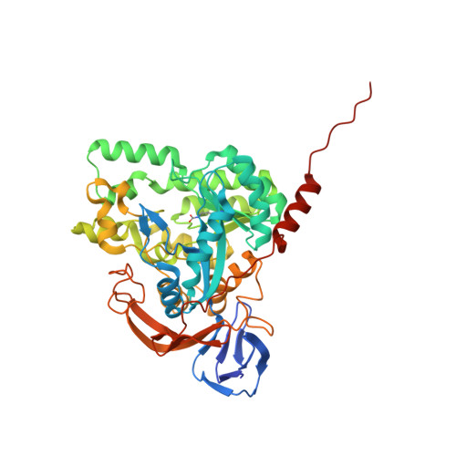 | |
UniProt | |||||
Find proteins for A0A0H3G9X2 (Brucella suis biovar 1 (strain 1330)) Explore A0A0H3G9X2 Go to UniProtKB: A0A0H3G9X2 | |||||
Entity Groups | |||||
| Sequence Clusters | 30% Identity50% Identity70% Identity90% Identity95% Identity100% Identity | ||||
| UniProt Group | A0A0H3G9X2 | ||||
Sequence AnnotationsExpand | |||||
Reference Sequence | |||||
| Ligands 2 Unique | |||||
|---|---|---|---|---|---|
| ID | Chains | Name / Formula / InChI Key | 2D Diagram | 3D Interactions | |
| ZN Download:Ideal Coordinates CCD File | EA [auth E] FA [auth E] G [auth A] H [auth A] KA [auth F] | ZINC ION Zn PTFCDOFLOPIGGS-UHFFFAOYSA-N |  | ||
| EDO Download:Ideal Coordinates CCD File | AA [auth D] BA [auth D] CA [auth D] DA [auth D] GA [auth E] | 1,2-ETHANEDIOL C2 H6 O2 LYCAIKOWRPUZTN-UHFFFAOYSA-N |  | ||
| Modified Residues 1 Unique | |||||
|---|---|---|---|---|---|
| ID | Chains | Type | Formula | 2D Diagram | Parent |
| KCX Query on KCX | A, B, C, D, E A, B, C, D, E, F | L-PEPTIDE LINKING | C7 H14 N2 O4 |  | LYS |
| Length ( Å ) | Angle ( ˚ ) |
|---|---|
| a = 156.69 | α = 90 |
| b = 88.83 | β = 91.17 |
| c = 221.24 | γ = 90 |
| Software Name | Purpose |
|---|---|
| XDS | data reduction |
| XSCALE | data scaling |
| PHASER | phasing |
| ARP | model building |
| PHENIX | refinement |
| PDB_EXTRACT | data extraction |


