4TPT | pdb_00004tpt

Crystal Structure of the Human LIMK2 Kinase Domain In Complex With a Non-ATP Competitive Inhibitor


Experimental Data Snapshot

  • Method: X-RAY DIFFRACTION
  • Resolution: 2.60 Å
  • R-Value Free: 
    0.274 (Depositor), 0.280 (DCC) 
  • R-Value Work: 
    0.220 (Depositor), 0.220 (DCC) 
  • R-Value Observed: 
    0.223 (Depositor) 

wwPDB Validation   3D Report Full Report


Ligand Structure Quality Assessment 

Created with Raphaël 2.3.0Worse 01 BetterLigand structure goodness of fit to experimental dataBest fitted 35HClick on this verticalbar to view details

This is version 2.1 of the entry. See complete history


Literature

Discovery of a Type III Inhibitor of LIM Kinase 2 That Binds in a DFG-Out Conformation.

Goodwin, N.C.Cianchetta, G.Burgoon, H.A.Healy, J.Mabon, R.Strobel, E.D.Allen, J.Wang, S.Hamman, B.D.Rawlins, D.B.

(2015) ACS Med Chem Lett 6: 53-57

  • DOI: https://doi.org/10.1021/ml500242y
  • Primary Citation of Related Structures:  
    4TPT

  • PubMed Abstract: 

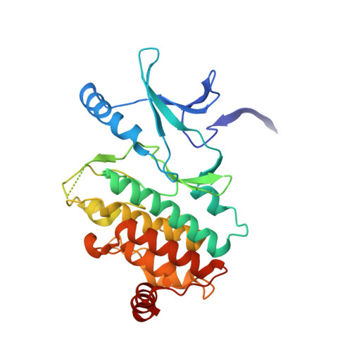
    The first allosteric, type III inhibitor of LIM-kinase 2 (LIMK2) is reported. A series of molecules that feature both an N-phenylsulfonamide and tertiary amide were not only very potent at LIMK2 but also were extremely selective against a panel of other kinases. Enzymatic kinetic studies showed these molecules to be noncompetitive with ATP, suggesting allosteric inhibition. X-ray crystallography confirmed that these sulfonamides are a rare example of a type III kinase inhibitor that binds away from the highly conserved hinge region and instead resides in the hydrophobic pocket formed in the DFG-out conformation of the kinase, thus accounting for the high level of selectivity observed.


  • Organizational Affiliation

    Lexicon Pharmaceuticals , 350 Carter Road, Princeton, New Jersey 08540, United States.


Macromolecules
Find similar proteins by:  (by identity cutoff)  |  3D Structure
Entity ID: 1
MoleculeChains Sequence LengthOrganismDetailsImage
LIM domain kinase 2
A, B
303Homo sapiensMutation(s): 0 
Gene Names: LIMK2
EC: 2.7.11.1
UniProt & NIH Common Fund Data Resources
Find proteins for P53671 (Homo sapiens)
Explore P53671 
Go to UniProtKB:  P53671
PHAROS:  P53671
GTEx:  ENSG00000182541 
Entity Groups  
Sequence Clusters30% Identity50% Identity70% Identity90% Identity95% Identity100% Identity
UniProt GroupP53671
Sequence Annotations
Expand
  • Reference Sequence
Small Molecules
Ligands 1 Unique
IDChains Name / Formula / InChI Key2D Diagram3D Interactions
35H
Query on 35H

Download Ideal Coordinates CCD File 
C [auth A],
D [auth B]
N-{4-[(1S)-1,2-dihydroxyethyl]benzyl}-N-methyl-4-(phenylsulfamoyl)benzamide
C23 H24 N2 O5 S
LJBXWGVECYNLLM-JOCHJYFZSA-N
Binding Affinity Annotations 
IDSourceBinding Affinity
35H BindingDB:  4TPT IC50: 92 (nM) from 1 assay(s)
Experimental Data & Validation

Experimental Data

  • Method: X-RAY DIFFRACTION
  • Resolution: 2.60 Å
  • R-Value Free:  0.274 (Depositor), 0.280 (DCC) 
  • R-Value Work:  0.220 (Depositor), 0.220 (DCC) 
  • R-Value Observed: 0.223 (Depositor) 
Space Group: P 1 21 1
Unit Cell:
Length ( Å )Angle ( ˚ )
a = 51.569α = 90
b = 77.903β = 100.84
c = 86.387γ = 90
Software Package:
Software NamePurpose
REFMACrefinement

Structure Validation

View Full Validation Report


Ligand Structure Quality Assessment 

Created with Raphaël 2.3.0Worse 01 BetterLigand structure goodness of fit to experimental dataBest fitted 35HClick on this verticalbar to view details

Entry History 

Deposition Data

Revision History  (Full details and data files)

  • Version 1.0: 2014-10-22
    Type: Initial release
  • Version 1.1: 2015-01-28
    Changes: Database references
  • Version 2.0: 2023-12-27
    Changes: Atomic model, Data collection, Database references, Derived calculations, Other, Refinement description, Source and taxonomy, Structure summary
  • Version 2.1: 2024-10-23
    Changes: Structure summary