4TL1 | pdb_00004tl1

GCN4-p1 with mutation to 1-Aminocyclohexanecarboxylic acid at residue 10


Experimental Data Snapshot

  • Method: X-RAY DIFFRACTION
  • Resolution: 1.80 Å
  • R-Value Free: 
    0.228 (Depositor), 0.241 (DCC) 
  • R-Value Work: 
    0.185 (Depositor), 0.196 (DCC) 
  • R-Value Observed: 
    0.187 (Depositor) 

Starting Model: experimental
View more details

wwPDB Validation 3D Report Full Report

Validation slider image for 4TL1

This is version 1.7 of the entry. See complete history

Literature

Origins of Structural Flexibility in Protein-Based Supramolecular Polymers Revealed by DEER Spectroscopy.

Tavenor, N.A.Silva, K.I.Saxena, S.Horne, W.S.

(2014) J Phys Chem B 118: 9881-9889

  • DOI: https://doi.org/10.1021/jp505643w
  • Primary Citation Related Structures: 
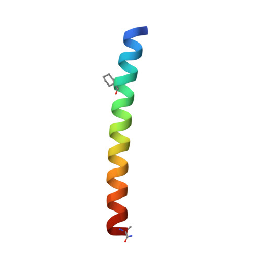
    4TL1

  • PubMed Abstract: 

    Modular assembly of bio-inspired supramolecular polymers is a powerful technique to develop new soft nanomaterials, and protein folding is a versatile basis for preparing such materials. Previous work demonstrated a significant difference in the physical properties of closely related supramolecular polymers composed of building blocks in which identical coiled-coil-forming peptides are cross-linked by one of two subtly different organic linkers (one flexible and the other rigid). Herein, we investigate the molecular basis for this observation by isolating a single subunit of the supramolecular polymer chain and probing its structure and conformational flexibility by double electron-electron resonance (DEER) spectroscopy. Experimental spin-spin distance distributions for two different labeling sites coupled with molecular dynamics simulations provide insights into how the linker structure impacts chain dynamics in the coiled-coil supramolecular polymer.


  • Organizational Affiliation
    • Department of Chemistry, University of Pittsburgh , Pittsburgh, Pennsylvania 15260, United States.

Macromolecule Content 

  • Total Structure Weight: 8.11 kDa 
  • Atom Count: 642 
  • Modeled Residue Count: 69 
  • Deposited Residue Count: 70 
  • Unique protein chains: 1

Macromolecules

Find similar proteins by:|  3D Structure
Entity ID: 1
MoleculeChains  Sequence LengthOrganismDetailsImage
General control protein GCN4
A, B
35Saccharomyces cerevisiae S288CMutation(s): 1 
UniProt
Find proteins for P03069 (Saccharomyces cerevisiae (strain ATCC 204508 / S288c))
Explore P03069 
Go to UniProtKB:  P03069
Entity Groups
Sequence Clusters30% Identity50% Identity70% Identity90% Identity95% Identity100% Identity
UniProt GroupP03069
Sequence Annotations
Expand
Reference Sequence

Small Molecules

Ligands 1 Unique
IDChains Name / Formula / InChI Key2D Diagram3D Interactions
IPA

Query on IPA


Download:Ideal Coordinates CCD File
C [auth A]ISOPROPYL ALCOHOL
C3 H8 O
KFZMGEQAYNKOFK-UHFFFAOYSA-N
Modified Residues  1 Unique
IDChains TypeFormula2D DiagramParent
02K
Query on 02K
A, B
PEPTIDE LINKINGC7 H13 N O2ALA

Experimental Data & Validation

Experimental Data

  • Method: X-RAY DIFFRACTION
  • Resolution: 1.80 Å
  • R-Value Free:  0.228 (Depositor), 0.241 (DCC) 
  • R-Value Work:  0.185 (Depositor), 0.196 (DCC) 
  • R-Value Observed: 0.187 (Depositor) 
Space Group: C 1 2 1
Unit Cell:
Length ( Å )Angle ( ˚ )
a = 83.322α = 90
b = 30.35β = 101.68
c = 28.019γ = 90
Software Package:
Software NamePurpose
CrystalCleardata collection
CrystalClearphasing
PHENIXphasing
PHENIXrefinement

Structure Validation

View Full Validation Report


Entry History 

& Funding Information

Deposition Data


Funding OrganizationLocationGrant Number
National Science Foundation (NSF, United States)United StatesDMR1149067

Revision History  (Full details and data files)

  • Version 1.0: 2014-08-06
    Type: Initial release
  • Version 1.1: 2014-08-20
    Changes: Structure summary
  • Version 1.2: 2014-10-01
    Changes: Database references
  • Version 1.3: 2017-09-13
    Changes: Author supporting evidence, Database references, Derived calculations, Other, Source and taxonomy, Structure summary
  • Version 1.4: 2017-11-22
    Changes: Refinement description
  • Version 1.5: 2019-11-27
    Changes: Author supporting evidence
  • Version 1.6: 2023-09-27
    Changes: Data collection, Database references, Refinement description
  • Version 1.7: 2024-11-20
    Changes: Structure summary