4RZB | pdb_00004rzb

The structure of N-formimino-L-Glutamate Iminohydrolase from Pseudomonas aeruginosa complexed with N-formimino-L-Aspartate, SOAKED WITH MERCURY


Experimental Data Snapshot

  • Method: X-RAY DIFFRACTION
  • Resolution: 1.86 Å
  • R-Value Free: 
    0.176 (Depositor), 0.177 (DCC) 
  • R-Value Work: 
    0.160 (Depositor), 0.161 (DCC) 
  • R-Value Observed: 
    0.160 (Depositor) 

wwPDB Validation 3D Report Full Report

Validation slider image for 4RZB

Ligand Structure Quality Assessment 


This is version 1.2 of the entry. See complete history

Literature

Structure of N-Formimino-l-glutamate Iminohydrolase from Pseudomonas aeruginosa.

Fedorov, A.A.Marti-Arbona, R.Nemmara, V.V.Hitchcock, D.Fedorov, E.V.Almo, S.C.Raushel, F.M.

(2015) Biochemistry 54: 890-897

  • DOI: https://doi.org/10.1021/bi501299y
  • Primary Citation Related Structures: 
    3MDU, 3MDW, 4RZB

  • PubMed Abstract: 

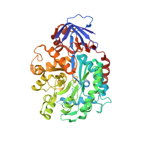
    N-Formimino-l-glutamate iminohydrolase (HutF), from Pseudomonas aeruginosa with a locus tag of Pa5106 ( gi|15600299 ), is a member of the amidohydrolase superfamily. This enzyme catalyzes the deamination of N-formimino-l-glutamate to N-formyl-l-glutamate and ammonia in the histidine degradation pathway. The crystal structure of Pa5106 was determined in the presence of the inhibitors N-formimino-l-aspartate and N-guanidino-l-glutaric acid at resolutions of 1.9 and 1.4 Å, respectively. The structure of an individual subunit is composed of two domains with the larger domain folding as a distorted (β/α)8-barrel. The (β/α)8-barrel domain is composed of eight β-strands flanked by 11 α-helices, whereas the smaller domain is made up of eight β-strands. The active site of Pa5106 contains a single zinc atom that is coordinated by His-56, His-58, His-232, and Asp-320. The nucleophilic solvent water molecule coordinates with the zinc atom at a distance of 2.0 Å and is hydrogen bonded to Asp-320 and His-269. The α-carboxylate groups of both inhibitors are hydrogen bonded to the imidazole moiety of His-206, the hydroxyl group of Tyr-121, and the side chain amide group of Gln-61. The side chain carboxylate groups of the two inhibitors are ion-paired with the guanidino groups of Arg-209 and Arg-82. Computational docking of high-energy tetrahedral intermediate forms of the substrate, N-formimino-l-glutamate, to the three-dimensional structure of Pa5106 suggests that this compound likely undergoes a re-faced nucleophilic attack at the formimino group by the metal-bound hydroxide. A catalytic mechanism of the reaction catalyzed by Pa5106 is proposed.


  • Organizational Affiliation
    • Department of Chemistry and ‡Department of Biochemistry & Biophysics, Texas A&M University , College Station, Texas 77843, United States.

Macromolecule Content 

  • Total Structure Weight: 100.94 kDa 
  • Atom Count: 7,971 
  • Modeled Residue Count: 902 
  • Deposited Residue Count: 906 
  • Unique protein chains: 1

Macromolecules

Find similar proteins by:|  3D Structure
Entity ID: 1
MoleculeChains  Sequence LengthOrganismDetailsImage
N-formimino-L-Glutamate Iminohydrolase
A, B
453Pseudomonas aeruginosa PAO1Mutation(s): 0 
Gene Names: PA5106
EC: 3.5.3.13
UniProt
Find proteins for Q9HU77 (Pseudomonas aeruginosa (strain ATCC 15692 / DSM 22644 / CIP 104116 / JCM 14847 / LMG 12228 / 1C / PRS 101 / PAO1))
Explore Q9HU77 
Go to UniProtKB:  Q9HU77
Entity Groups
Sequence Clusters30% Identity50% Identity70% Identity90% Identity95% Identity100% Identity
UniProt GroupQ9HU77
Sequence Annotations
Expand
Reference Sequence

Small Molecules

Ligands 5 Unique
IDChains Name / Formula / InChI Key2D Diagram3D Interactions
HG

Query on HG


Download:Ideal Coordinates CCD File
E [auth A]
F [auth A]
G [auth A]
L [auth B]
M [auth B]
E [auth A],
F [auth A],
G [auth A],
L [auth B],
M [auth B],
N [auth B]
MERCURY (II) ION
Hg
BQPIGGFYSBELGY-UHFFFAOYSA-N
NFQ

Query on NFQ


Download:Ideal Coordinates CCD File
C [auth A],
J [auth B]
N-[(E)-iminomethyl]-L-aspartic acid
C5 H8 N2 O4
XTPIFIMCFHNJOH-VKHMYHEASA-N
SO4

Query on SO4


Download:Ideal Coordinates CCD File
H [auth A],
O [auth B],
P [auth B],
Q [auth B]
SULFATE ION
O4 S
QAOWNCQODCNURD-UHFFFAOYSA-L
GOL

Query on GOL


Download:Ideal Coordinates CCD File
I [auth A]GLYCEROL
C3 H8 O3
PEDCQBHIVMGVHV-UHFFFAOYSA-N
ZN

Query on ZN


Download:Ideal Coordinates CCD File
D [auth A],
K [auth B]
ZINC ION
Zn
PTFCDOFLOPIGGS-UHFFFAOYSA-N

Experimental Data & Validation

Experimental Data

  • Method: X-RAY DIFFRACTION
  • Resolution: 1.86 Å
  • R-Value Free:  0.176 (Depositor), 0.177 (DCC) 
  • R-Value Work:  0.160 (Depositor), 0.161 (DCC) 
  • R-Value Observed: 0.160 (Depositor) 
Space Group: C 1 2 1
Unit Cell:
Length ( Å )Angle ( ˚ )
a = 101.56α = 90
b = 141.831β = 107.18
c = 86.415γ = 90
Software Package:
Software NamePurpose
ADSCdata collection
PHENIXmodel building
PHENIXrefinement
HKL-2000data reduction
HKL-2000data scaling
PHENIXphasing

Structure Validation

View Full Validation Report


Ligand Structure Quality Assessment 


Entry History 

Deposition Data

Revision History  (Full details and data files)

  • Version 1.0: 2015-01-14
    Type: Initial release
  • Version 1.1: 2015-02-11
    Changes: Database references
  • Version 1.2: 2024-02-28
    Changes: Data collection, Database references, Derived calculations