4RWR | pdb_00004rwr

2.1 Angstrom Crystal Structure of Stage II Sporulation Protein D from Bacillus anthracis


Experimental Data Snapshot

  • Method: X-RAY DIFFRACTION
  • Resolution: 2.10 Å
  • R-Value Free: 
    0.243 (Depositor), 0.249 (DCC) 
  • R-Value Work: 
    0.188 (Depositor), 0.196 (DCC) 
  • R-Value Observed: 
    0.190 (Depositor) 

wwPDB Validation 3D Report Full Report

Validation slider image for 4RWR

This is version 1.4 of the entry. See complete history

Literature

Crystal Structures of the SpoIID Lytic Transglycosylases Essential for Bacterial Sporulation.

Nocadello, S.Minasov, G.Shuvalova, L.S.Dubrovska, I.Sabini, E.Anderson, W.F.

(2016) J Biological Chem 291: 14915-14926

  • DOI: https://doi.org/10.1074/jbc.M116.729749
  • Primary Citation Related Structures: 
    4RWR, 5I1T

  • PubMed Abstract: 

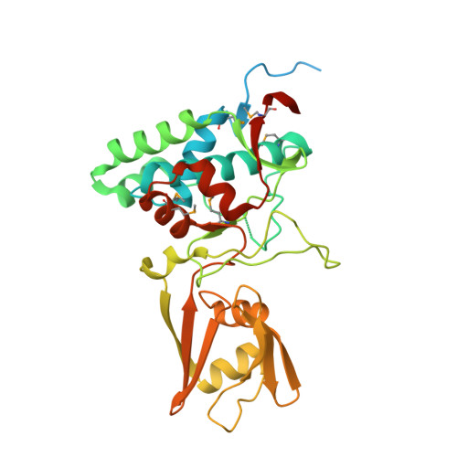
    Bacterial spores are the most resistant form of life known on Earth and represent a serious problem for (i) bioterrorism attack, (ii) horizontal transmission of microbial pathogens in the community, and (iii) persistence in patients and in a nosocomial environment. Stage II sporulation protein D (SpoIID) is a lytic transglycosylase (LT) essential for sporulation. The LT superfamily is a potential drug target because it is active in essential bacterial processes involving the peptidoglycan, which is unique to bacteria. However, the absence of structural information for the sporulation-specific LT enzymes has hindered mechanistic understanding of SpoIID. Here, we report the first crystal structures with and without ligands of the SpoIID family from two community relevant spore-forming pathogens, Bacillus anthracis and Clostridium difficile. The structures allow us to visualize the overall architecture, characterize the substrate recognition model, identify critical residues, and provide the structural basis for catalysis by this new family of enzymes.


  • Organizational Affiliation
    • From the Center for Structural Genomics of Infectious Diseases, Department of Biochemistry and Molecular Genetics, Northwestern University Feinberg School of Medicine, Chicago, Illinois 60611.

Macromolecule Content 

  • Total Structure Weight: 74.11 kDa 
  • Atom Count: 4,642 
  • Modeled Residue Count: 535 
  • Deposited Residue Count: 662 
  • Unique protein chains: 1

Macromolecules

Find similar proteins by:|  3D Structure
Entity ID: 1
MoleculeChains  Sequence LengthOrganismDetailsImage
Stage II sporulation protein D
A, B
331Bacillus anthracisMutation(s): 0 
Gene Names: BAS5136BA_5528DH23_5378DJ42_3419DJ44_3608DJ45_519DJ48_4453DJ49_4581GBAA_5528spoIID
UniProt
Find proteins for A0A6L7HNW1 (Bacillus anthracis)
Explore A0A6L7HNW1 
Go to UniProtKB:  A0A6L7HNW1
Entity Groups
Sequence Clusters30% Identity50% Identity70% Identity90% Identity95% Identity100% Identity
UniProt GroupA0A6L7HNW1
Sequence Annotations
Expand
Reference Sequence

Small Molecules

Modified Residues  1 Unique
IDChains TypeFormula2D DiagramParent
MSE
Query on MSE
A, B
L-PEPTIDE LINKINGC5 H11 N O2 SeMET

Experimental Data & Validation

Experimental Data

  • Method: X-RAY DIFFRACTION
  • Resolution: 2.10 Å
  • R-Value Free:  0.243 (Depositor), 0.249 (DCC) 
  • R-Value Work:  0.188 (Depositor), 0.196 (DCC) 
  • R-Value Observed: 0.190 (Depositor) 
Space Group: P 1 21 1
Unit Cell:
Length ( Å )Angle ( ˚ )
a = 46.616α = 90
b = 144.477β = 115.36
c = 47.076γ = 90
Software Package:
Software NamePurpose
Blu-Icedata collection
PHENIXmodel building
REFMACrefinement
HKL-3000data reduction
HKL-3000data scaling
PHENIXphasing

Structure Validation

View Full Validation Report


Entry History 

Revision History  (Full details and data files)

  • Version 1.0: 2014-12-17
    Type: Initial release
  • Version 1.1: 2016-06-08
    Changes: Database references
  • Version 1.2: 2016-08-03
    Changes: Database references
  • Version 1.3: 2017-11-22
    Changes: Refinement description
  • Version 1.4: 2024-11-06
    Changes: Data collection, Database references, Derived calculations, Structure summary