4RW3 | pdb_00004rw3

Structural insights into substrate binding of brown spider venom class II phospholipases D


Experimental Data Snapshot

  • Method: X-RAY DIFFRACTION
  • Resolution: 1.72 Å
  • R-Value Free: 
    0.223 (Depositor), 0.223 (DCC) 
  • R-Value Work: 
    0.183 (Depositor), 0.185 (DCC) 
  • R-Value Observed: 
    0.187 (Depositor) 

Starting Model: experimental
View more details

wwPDB Validation 3D Report Full Report

Validation slider image for 4RW3

Ligand Structure Quality Assessment 


This is version 1.3 of the entry. See complete history

Literature

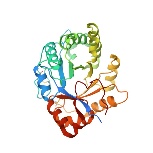
Structural Insights into Substrate Binding of Brown Spider Venom Class II Phospholipases D.

Coronado, M.A.Ullah, A.da Silva, L.S.Chaves-Moreira, D.Vuitika, L.Chaim, O.M.Veiga, S.S.Chahine, J.Murakami, M.T.Arni, R.K.

(2015) Curr Protein Pept Sci 16: 768-774

  • DOI: https://doi.org/10.2174/1389203716666150505231625
  • Primary Citation Related Structures: 
    4RW3, 4RW5

  • PubMed Abstract: 

    Phospholipases D (PLDs), the major dermonecrotic factors from brown spider venoms, trigger a range of biological reactions both in vitro and in vivo. Despite their clinical relevance in loxoscelism, structural data is restricted to the apo-form of these enzymes, which has been instrumental in understanding the functional differences between the class I and II spider PLDs. The crystal structures of the native class II PLD from Loxosceles intermedia complexed with myo-inositol 1-phosphate and the inactive mutant H12A complexed with fatty acids indicate the existence of a strong ligand-dependent conformation change of the highly conserved aromatic residues, Tyr 223 and Trp225 indicating their roles in substrate binding. These results provided insights into the structural determinants for substrate recognition and binding by class II PLDs.


  • Organizational Affiliation
    • Centro Multiusuario de Inovacao Biomolecular, Departamento de Fisica, Universidade Estadual Paulista (UNESP), Sao Jose do Rio Preto, 15054-000 SP, Brazil. arni@sjrp.unesp.br.

Macromolecule Content 

  • Total Structure Weight: 35.93 kDa 
  • Atom Count: 2,558 
  • Modeled Residue Count: 279 
  • Deposited Residue Count: 302 
  • Unique protein chains: 1

Macromolecules

Find similar proteins by:|  3D Structure
Entity ID: 1
MoleculeChains  Sequence LengthOrganismDetailsImage
Phospholipase D LiSicTox-alphaIA1bii302Loxosceles intermediaMutation(s): 0 
EC: 3.1.4.4 (PDB Primary Data), 4.6.1 (UniProt)
UniProt
Find proteins for P0CE82 (Loxosceles intermedia)
Explore P0CE82 
Go to UniProtKB:  P0CE82
Entity Groups
Sequence Clusters30% Identity50% Identity70% Identity90% Identity95% Identity100% Identity
UniProt GroupP0CE82
Sequence Annotations
Expand
Reference Sequence

Small Molecules

Ligands 9 Unique
IDChains Name / Formula / InChI Key2D Diagram3D Interactions
IPD

Query on IPD


Download:Ideal Coordinates CCD File
E [auth A]D-MYO-INOSITOL-1-PHOSPHATE
C6 H11 O9 P
INAPMGSXUVUWAF-UOTPTPDRSA-L
PLM

Query on PLM


Download:Ideal Coordinates CCD File
J [auth A],
K [auth A],
L [auth A]
PALMITIC ACID
C16 H32 O2
IPCSVZSSVZVIGE-UHFFFAOYSA-N
TDA

Query on TDA


Download:Ideal Coordinates CCD File
N [auth A]N-TRIDECANOIC ACID
C13 H26 O2
SZHOJFHSIKHZHA-UHFFFAOYSA-N
DKA

Query on DKA


Download:Ideal Coordinates CCD File
I [auth A],
M [auth A]
DECANOIC ACID
C10 H20 O2
GHVNFZFCNZKVNT-UHFFFAOYSA-N
OCA

Query on OCA


Download:Ideal Coordinates CCD File
G [auth A]OCTANOIC ACID (CAPRYLIC ACID)
C8 H16 O2
WWZKQHOCKIZLMA-UHFFFAOYSA-N
SHV

Query on SHV


Download:Ideal Coordinates CCD File
H [auth A]HEPTANOIC ACID
C7 H14 O2
MNWFXJYAOYHMED-UHFFFAOYSA-N
PEG

Query on PEG


Download:Ideal Coordinates CCD File
C [auth A],
D [auth A]
DI(HYDROXYETHYL)ETHER
C4 H10 O3
MTHSVFCYNBDYFN-UHFFFAOYSA-N
GOL

Query on GOL


Download:Ideal Coordinates CCD File
F [auth A]GLYCEROL
C3 H8 O3
PEDCQBHIVMGVHV-UHFFFAOYSA-N
MG

Query on MG


Download:Ideal Coordinates CCD File
B [auth A]MAGNESIUM ION
Mg
JLVVSXFLKOJNIY-UHFFFAOYSA-N

Experimental Data & Validation

Experimental Data

  • Method: X-RAY DIFFRACTION
  • Resolution: 1.72 Å
  • R-Value Free:  0.223 (Depositor), 0.223 (DCC) 
  • R-Value Work:  0.183 (Depositor), 0.185 (DCC) 
  • R-Value Observed: 0.187 (Depositor) 
Space Group: P 1 21 1
Unit Cell:
Length ( Å )Angle ( ˚ )
a = 49.81α = 90
b = 49.3β = 105.83
c = 56.3γ = 90
Software Package:
Software NamePurpose
HKL-2000data collection
MOLREPphasing
REFMACrefinement
HKL-2000data reduction
SCALEPACKdata scaling

Structure Validation

View Full Validation Report


Ligand Structure Quality Assessment 


Entry History 

Deposition Data

Revision History  (Full details and data files)

  • Version 1.0: 2015-06-03
    Type: Initial release
  • Version 1.1: 2015-12-16
    Changes: Database references
  • Version 1.2: 2023-09-20
    Changes: Data collection, Database references, Derived calculations, Refinement description
  • Version 1.3: 2024-10-16
    Changes: Structure summary