


Crystal structure of a Putative Acyl-CoA ligase (BT_0428) from Bacteroides thetaiotaomicron VPI-5482 at 2.41 A resolution
Joint Center for Structural Genomics (JCSG)To be published.
Experimental Data Snapshot
Entity ID: 1 | |||||
|---|---|---|---|---|---|
| Molecule | Chains | Sequence Length | Organism | Details | Image |
| Phenylacetate-coenzyme A ligase | 436 | Bacteroides thetaiotaomicron VPI-5482 | Mutation(s): 0 Gene Names: BT_0428 EC: 6.2.1.30 | 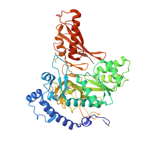 | |
UniProt | |||||
Entity Groups | |||||
| Sequence Clusters | 30% Identity50% Identity70% Identity90% Identity95% Identity100% Identity | ||||
| UniProt Group | Q8AAN6 | ||||
Sequence AnnotationsExpand | |||||
Reference Sequence | |||||
| Ligands 9 Unique | |||||
|---|---|---|---|---|---|
| ID | Chains | Name / Formula / InChI Key | 2D Diagram | 3D Interactions | |
| ADP Download:Ideal Coordinates CCD File | F [auth A], FA [auth C], OA [auth D], W [auth B] | ADENOSINE-5'-DIPHOSPHATE C10 H15 N5 O10 P2 XTWYTFMLZFPYCI-KQYNXXCUSA-N |  | ||
| PG4 Download:Ideal Coordinates CCD File | N [auth A] | TETRAETHYLENE GLYCOL C8 H18 O5 UWHCKJMYHZGTIT-UHFFFAOYSA-N |  | ||
| PGE Download:Ideal Coordinates CCD File | KA [auth C], P [auth A] | TRIETHYLENE GLYCOL C6 H14 O4 ZIBGPFATKBEMQZ-UHFFFAOYSA-N |  | ||
| PEG Download:Ideal Coordinates CCD File | AA [auth B], O [auth A] | DI(HYDROXYETHYL)ETHER C4 H10 O3 MTHSVFCYNBDYFN-UHFFFAOYSA-N |  | ||
| SO4 Download:Ideal Coordinates CCD File | JA [auth C] K [auth A] L [auth A] M [auth A] SA [auth D] | SULFATE ION O4 S QAOWNCQODCNURD-UHFFFAOYSA-L |  | ||
| ZN Download:Ideal Coordinates CCD File | E [auth A], EA [auth C], PA [auth D], V [auth B] | ZINC ION Zn PTFCDOFLOPIGGS-UHFFFAOYSA-N |  | ||
| EDO Download:Ideal Coordinates CCD File | BA [auth B] CA [auth B] DA [auth B] LA [auth C] MA [auth C] | 1,2-ETHANEDIOL C2 H6 O2 LYCAIKOWRPUZTN-UHFFFAOYSA-N |  | ||
| K Download:Ideal Coordinates CCD File | G [auth A] GA [auth C] H [auth A] HA [auth C] I [auth A] | POTASSIUM ION K NPYPAHLBTDXSSS-UHFFFAOYSA-N |  | ||
| UNL Download:Ideal Coordinates CCD File | IA [auth C], J [auth A], RA [auth D], X [auth B] | Unknown ligand NPYPAHLBTDXSSS-UHFFFAOYSA-N | |||
| Modified Residues 1 Unique | |||||
|---|---|---|---|---|---|
| ID | Chains | Type | Formula | 2D Diagram | Parent |
| MSE Query on MSE | A, B, C, D | L-PEPTIDE LINKING | C5 H11 N O2 Se |  | MET |
| Length ( Å ) | Angle ( ˚ ) |
|---|---|
| a = 127.237 | α = 90 |
| b = 210.223 | β = 90 |
| c = 71.504 | γ = 90 |
| Software Name | Purpose |
|---|---|
| MolProbity | model building |
| PDB_EXTRACT | data extraction |
| SHELX | phasing |
| SHARP | phasing |
| SCALA | data scaling |
| BUSTER-TNT | refinement |
| XDS | data reduction |
| SHELXD | phasing |
| autoSHARP | phasing |
| BUSTER | refinement |


