4RSU

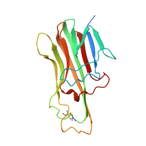
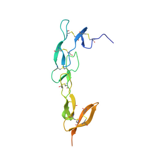
Crystal structure of the light and hvem complex


Experimental Data Snapshot

  • Method: X-RAY DIFFRACTION
  • Resolution: 2.30 Å
  • R-Value Free: 0.231 
  • R-Value Work: 0.188 
  • R-Value Observed: 0.191 

Starting Models: experimental
View more details

wwPDB Validation   3D Report Full Report


Ligand Structure Quality Assessment 


This is version 1.5 of the entry. See complete history


Literature

HVEM structures and mutants reveal distinct functions of binding to LIGHT and BTLA/CD160.

Liu, W.Chou, T.F.Garrett-Thomson, S.C.Seo, G.Y.Fedorov, E.Ramagopal, U.A.Bonanno, J.B.Wang, Q.Kim, K.Garforth, S.J.Kakugawa, K.Cheroutre, H.Kronenberg, M.Almo, S.C.

(2021) J Exp Med 218

  • DOI: https://doi.org/10.1084/jem.20211112
  • Primary Citation of Related Structures:  
    4RSU, 7MSG, 7MSJ

  • PubMed Abstract: 

    HVEM is a TNF (tumor necrosis factor) receptor contributing to a broad range of immune functions involving diverse cell types. It interacts with a TNF ligand, LIGHT, and immunoglobulin (Ig) superfamily members BTLA and CD160. Assessing the functional impact of HVEM binding to specific ligands in different settings has been complicated by the multiple interactions of HVEM and HVEM binding partners. To dissect the molecular basis for multiple functions, we determined crystal structures that reveal the distinct HVEM surfaces that engage LIGHT or BTLA/CD160, including the human HVEM-LIGHT-CD160 ternary complex, with HVEM interacting simultaneously with both binding partners. Based on these structures, we generated mouse HVEM mutants that selectively recognized either the TNF or Ig ligands in vitro. Knockin mice expressing these muteins maintain expression of all the proteins in the HVEM network, yet they demonstrate selective functions for LIGHT in the clearance of bacteria in the intestine and for the Ig ligands in the amelioration of liver inflammation.


  • Organizational Affiliation

    Department of Biochemistry, Albert Einstein College of Medicine, Bronx, NY.


Macromolecules
Find similar proteins by:  (by identity cutoff)  |  3D Structure
Entity ID: 1
MoleculeChains Sequence LengthOrganismDetailsImage
Tumor necrosis factor ligand superfamily member 14, soluble form
A, B, C, G, H
A, B, C, G, H, I
165Homo sapiensMutation(s): 0 
Gene Names: TNFSF14HVEMLLIGHTUNQ391/PRO726
UniProt & NIH Common Fund Data Resources
Find proteins for O43557 (Homo sapiens)
Explore O43557 
Go to UniProtKB:  O43557
PHAROS:  O43557
GTEx:  ENSG00000125735 
Entity Groups  
Sequence Clusters30% Identity50% Identity70% Identity90% Identity95% Identity100% Identity
UniProt GroupO43557
Sequence Annotations
Expand
  • Reference Sequence
Find similar proteins by:  (by identity cutoff)  |  3D Structure
Entity ID: 2
MoleculeChains Sequence LengthOrganismDetailsImage
Tumor necrosis factor receptor superfamily member 14
D, E, F, J, K
D, E, F, J, K, L
134Homo sapiensMutation(s): 0 
Gene Names: TNFRSF14HVEAHVEMUNQ329/PRO509
UniProt & NIH Common Fund Data Resources
Find proteins for Q92956 (Homo sapiens)
Explore Q92956 
Go to UniProtKB:  Q92956
PHAROS:  Q92956
GTEx:  ENSG00000157873 
Entity Groups  
Sequence Clusters30% Identity50% Identity70% Identity90% Identity95% Identity100% Identity
UniProt GroupQ92956
Glycosylation
Glycosylation Sites: 1Go to GlyGen: Q92956-1
Sequence Annotations
Expand
  • Reference Sequence
Small Molecules
Ligands 3 Unique
IDChains Name / Formula / InChI Key2D Diagram3D Interactions
NAG
Query on NAG

Download Ideal Coordinates CCD File 
IA [auth J],
KA [auth K],
MA [auth L],
V [auth D],
X [auth E]
2-acetamido-2-deoxy-beta-D-glucopyranose
C8 H15 N O6
OVRNDRQMDRJTHS-FMDGEEDCSA-N
GOL
Query on GOL

Download Ideal Coordinates CCD File 
BA [auth G]
EA [auth H]
GA [auth I]
HA [auth I]
Q [auth B]
BA [auth G],
EA [auth H],
GA [auth I],
HA [auth I],
Q [auth B],
S [auth C],
U [auth D],
Y [auth F]
GLYCEROL
C3 H8 O3
PEDCQBHIVMGVHV-UHFFFAOYSA-N
CL
Query on CL

Download Ideal Coordinates CCD File 
AA [auth G]
CA [auth G]
DA [auth H]
FA [auth I]
JA [auth K]
AA [auth G],
CA [auth G],
DA [auth H],
FA [auth I],
JA [auth K],
LA [auth L],
M [auth A],
N [auth A],
O [auth B],
P [auth B],
R [auth C],
T [auth C],
W [auth E],
Z [auth G]
CHLORIDE ION
Cl
VEXZGXHMUGYJMC-UHFFFAOYSA-M
Experimental Data & Validation

Experimental Data

  • Method: X-RAY DIFFRACTION
  • Resolution: 2.30 Å
  • R-Value Free: 0.231 
  • R-Value Work: 0.188 
  • R-Value Observed: 0.191 
  • Space Group: P 21 21 21
Unit Cell:
Length ( Å )Angle ( ˚ )
a = 111.651α = 90
b = 113.604β = 90
c = 163.279γ = 90
Software Package:
Software NamePurpose
MOLREPphasing
REFMACrefinement
HKL-2000data reduction
HKL-2000data scaling

Structure Validation

View Full Validation Report


Ligand Structure Quality Assessment 


Entry History 

Revision History  (Full details and data files)

  • Version 1.0: 2015-02-04
    Type: Initial release
  • Version 1.1: 2015-02-11
    Changes: Structure summary
  • Version 1.2: 2020-07-29
    Type: Remediation
    Reason: Carbohydrate remediation
    Changes: Data collection, Database references, Derived calculations, Structure summary
  • Version 1.3: 2022-04-27
    Changes: Database references, Structure summary
  • Version 1.4: 2023-09-20
    Changes: Data collection, Refinement description
  • Version 1.5: 2024-11-20
    Changes: Structure summary