4RO1 | pdb_00004ro1

An 3'-5'-exoribonuclease that specifically recognizes RNAs.


Experimental Data Snapshot

  • Method: X-RAY DIFFRACTION
  • Resolution: 2.80 Å
  • R-Value Free: 
    0.294 (Depositor), 0.300 (DCC) 
  • R-Value Work: 
    0.269 (Depositor), 0.289 (DCC) 
  • R-Value Observed: 
    0.270 (Depositor) 

wwPDB Validation 3D Report Full Report

Validation slider image for 4RO1

This is version 1.2 of the entry. See complete history

Literature

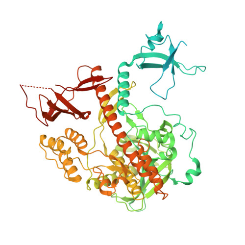
Structural analysis of Dis3l2, an exosome-independent exonuclease from Schizosaccharomyces pombe.

Lv, H.Zhu, Y.Qiu, Y.Niu, L.Teng, M.Li, X.

(2015) Acta Crystallogr D Biol Crystallogr 71: 1284-1294

  • DOI: https://doi.org/10.1107/S1399004715005805
  • Primary Citation Related Structures: 
    4RO1

  • PubMed Abstract: 

    After deadenylation and decapping, cytoplasmic mRNA can be digested in two opposite directions: in the 5'-3' direction by Xrn1 or in the 3'-5' direction by the exosome complex. Recently, a novel 3'-5' RNA-decay pathway involving Dis3l2 has been described that differs from degradation by Xrn1 and the exosome. The product of the Schizosaccharomyces pombe gene SPAC2C4.07c was identified as a homologue of human Dis3l2. In this work, the 2.8 Å resolution X-ray crystal structure of S. pombe Dis3l2 (SpDis3l2) is reported, the conformation of which is obviously different from that in the homologous mouse Dis3l2-RNA complex. Fluorescence polarization assay experiments showed that RNB and S1 are the primary RNA-binding domains and that the CSDs (CSD1 and CSD2) play an indispensable role in the RNA-binding process of SpDis3l2. Taking the structure comparison and mutagenic experiments together, it can be inferred that the RNA-recognition pattern of SpDis3l2 resembles that of its mouse homologue rather than that of the Escherichia coli RNase II-RNA complex. Furthermore, a drastic conformation change could occur following the binding of the RNA substrate to SpDis3l2.


  • Organizational Affiliation
    • Hefei National Laboratory for Physical Sciences at Microscale and School of Life Science, University of Science and Technology of China, Hefei, Anhui 230026, People's Republic of China.

Macromolecule Content 

  • Total Structure Weight: 174.13 kDa 
  • Atom Count: 8,275 
  • Modeled Residue Count: 1,134 
  • Deposited Residue Count: 1,528 
  • Unique protein chains: 1

Macromolecules

Find similar proteins by:|  3D Structure
Entity ID: 1
MoleculeChains  Sequence LengthOrganismDetailsImage
DIS3-like exonuclease 2A [auth B],
B [auth A]
764Schizosaccharomyces pombe 972h-Mutation(s): 0 
Gene Names: dis32dis3l2SPAC2C4.07c
EC: 3.1.13
UniProt
Find proteins for O14040 (Schizosaccharomyces pombe (strain 972 / ATCC 24843))
Explore O14040 
Go to UniProtKB:  O14040
Entity Groups
Sequence Clusters30% Identity50% Identity70% Identity90% Identity95% Identity100% Identity
UniProt GroupO14040
Sequence Annotations
Expand
Reference Sequence

Experimental Data & Validation

Experimental Data

  • Method: X-RAY DIFFRACTION
  • Resolution: 2.80 Å
  • R-Value Free:  0.294 (Depositor), 0.300 (DCC) 
  • R-Value Work:  0.269 (Depositor), 0.289 (DCC) 
  • R-Value Observed: 0.270 (Depositor) 
Space Group: P 1 21 1
Unit Cell:
Length ( Å )Angle ( ˚ )
a = 116.966α = 90
b = 54.201β = 107.09
c = 127.217γ = 90
Software Package:
Software NamePurpose
HKL-2000data collection
AutoSolphasing
PHENIXrefinement
HKL-2000data reduction
HKL-2000data scaling

Structure Validation

View Full Validation Report


Entry History 

Deposition Data

Revision History  (Full details and data files)

  • Version 1.0: 2015-06-10
    Type: Initial release
  • Version 1.1: 2015-07-01
    Changes: Database references
  • Version 1.2: 2024-02-28
    Changes: Data collection, Database references