4RKD | pdb_00004rkd

Psychrophilic aromatic amino acids aminotransferase from Psychrobacter sp. B6 cocrystalized with aspartic acid


Experimental Data Snapshot

  • Method: X-RAY DIFFRACTION
  • Resolution: 2.76 Å
  • R-Value Free: 
    0.222 (Depositor), 0.220 (DCC) 
  • R-Value Work: 
    0.173 (Depositor), 0.173 (DCC) 
  • R-Value Observed: 
    0.176 (Depositor) 

Starting Model: experimental
View more details

wwPDB Validation 3D Report Full Report

Validation slider image for 4RKD

Ligand Structure Quality Assessment 


This is version 1.3 of the entry. See complete history

Literature

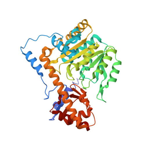
Crystal structure and enzymatic properties of a broad substrate-specificity psychrophilic aminotransferase from the Antarctic soil bacterium Psychrobacter sp. B6.

Bujacz, A.Rutkiewicz-Krotewicz, M.Nowakowska-Sapota, K.Turkiewicz, M.

(2015) Acta Crystallogr D Biol Crystallogr 71: 632-645

  • DOI: https://doi.org/10.1107/S1399004714028016
  • Primary Citation Related Structures: 
    4RKC, 4RKD

  • PubMed Abstract: 

    Aminotransferases (ATs) are enzymes that are commonly used in the chemical and pharmaceutical industries for the synthesis of natural and non-natural amino acids by transamination reactions. Currently, the easily accessible enzymes from mesophilic organisms are most commonly used; however, for economical and ecological reasons the utilization of aminotransferases from psychrophiles would be more advantageous, as their optimum reaction temperature is usually significantly lower than for the mesophilic ATs. Here, gene isolation, protein expression, purification, enzymatic properties and structural studies are reported for the cold-active aromatic amino-acid aminotransferase (PsyArAT) from Psychrobacter sp. B6, a psychrotrophic, Gram-negative strain from Antarctic soil. Preliminary computational analysis indicated dual functionality of the enzyme through the ability to utilize both aromatic amino acids and aspartate as substrates. This postulation was confirmed by enzymatic activity tests, which showed that it belonged to the class EC 2.6.1.57. The first crystal structures of a psychrophilic aromatic amino-acid aminotransferase have been determined at resolutions of 2.19 Å for the native enzyme (PsyArAT) and 2.76 Å for its complex with aspartic acid (PsyArAT/D). Both types of crystals grew in the monoclinic space group P21 under slightly different crystallization conditions. The PsyArAT crystals contained a dimer (90 kDa) in the asymmetric unit, which corresponds to the active form of this enzyme, whereas the crystals of the PsyArAT/D complex included four dimers showing different stages of the transamination reaction.


  • Organizational Affiliation
    • Institute of Technical Biochemistry, Lodz University of Technology, Stefanowskiego 4/10, 90-924 Lodz, Poland.

Macromolecule Content 

  • Total Structure Weight: 356.77 kDa 
  • Atom Count: 25,951 
  • Modeled Residue Count: 3,184 
  • Deposited Residue Count: 3,184 
  • Unique protein chains: 1

Macromolecules

Find similar proteins by:|  3D Structure
Entity ID: 1
MoleculeChains  Sequence LengthOrganismDetailsImage
Aromatic amino acid aminotransferase
A, B, C, D, E
A, B, C, D, E, F, G, H
398Psychrobacter sp. B6Mutation(s): 0 
EC: 2.6.1.57 (PDB Primary Data), 2.6.1 (UniProt)
UniProt
Find proteins for C7E5X4 (Psychrobacter sp. B6)
Explore C7E5X4 
Go to UniProtKB:  C7E5X4
Entity Groups
Sequence Clusters30% Identity50% Identity70% Identity90% Identity95% Identity100% Identity
UniProt GroupC7E5X4
Sequence Annotations
Expand
Reference Sequence

Small Molecules

Ligands 5 Unique
IDChains Name / Formula / InChI Key2D Diagram3D Interactions
KET

Query on KET


Download:Ideal Coordinates CCD File
K [auth A],
T [auth E],
W [auth G]
2-[(3-HYDROXY-2-METHYL-5-PHOSPHONOOXYMETHYL-PYRIDIN-4-YLMETHYLENE)-AMINO]-SUCCINIC ACID
C12 H16 N2 O9 P
OEULMZWKJBFRJF-LPQZUGQSSA-O
PMP

Query on PMP


Download:Ideal Coordinates CCD File
P [auth D],
X [auth H]
4'-DEOXY-4'-AMINOPYRIDOXAL-5'-PHOSPHATE
C8 H13 N2 O5 P
ZMJGSOSNSPKHNH-UHFFFAOYSA-N
PLP

Query on PLP


Download:Ideal Coordinates CCD File
J [auth A],
L [auth B],
N [auth C],
R [auth E],
U [auth F]
PYRIDOXAL-5'-PHOSPHATE
C8 H10 N O6 P
NGVDGCNFYWLIFO-UHFFFAOYSA-N
OAA

Query on OAA


Download:Ideal Coordinates CCD File
O [auth C],
S [auth E]
OXALOACETATE ION
C4 H3 O5
KHPXUQMNIQBQEV-UHFFFAOYSA-M
MG

Query on MG


Download:Ideal Coordinates CCD File
I [auth A],
M [auth C],
Q [auth E],
V [auth G]
MAGNESIUM ION
Mg
JLVVSXFLKOJNIY-UHFFFAOYSA-N

Experimental Data & Validation

Experimental Data

  • Method: X-RAY DIFFRACTION
  • Resolution: 2.76 Å
  • R-Value Free:  0.222 (Depositor), 0.220 (DCC) 
  • R-Value Work:  0.173 (Depositor), 0.173 (DCC) 
  • R-Value Observed: 0.176 (Depositor) 
Space Group: P 1 21 1
Unit Cell:
Length ( Å )Angle ( ˚ )
a = 92.25α = 90
b = 103.23β = 98.58
c = 165.78γ = 90
Software Package:
Software NamePurpose
HKL-2000data collection
MOLREPphasing
REFMACrefinement
XDSdata reduction
XDSdata scaling

Structure Validation

View Full Validation Report


Ligand Structure Quality Assessment 


Entry History 

Deposition Data

Revision History  (Full details and data files)

  • Version 1.0: 2015-03-11
    Type: Initial release
  • Version 1.1: 2015-03-18
    Changes: Database references
  • Version 1.2: 2015-04-15
    Changes: Database references
  • Version 1.3: 2023-09-20
    Changes: Data collection, Database references, Derived calculations, Refinement description