4R7P | pdb_00004r7p

Human UDP-glucose pyrophosphorylase isoform 1 in complex with UDP-glucose


Experimental Data Snapshot

  • Method: X-RAY DIFFRACTION
  • Resolution: 3.35 Å
  • R-Value Free: 
    0.247 (Depositor), 0.251 (DCC) 
  • R-Value Work: 
    0.199 (Depositor), 0.205 (DCC) 
  • R-Value Observed: 
    0.202 (Depositor) 

wwPDB Validation 3D Report Full Report

Validation slider image for 4R7P

Ligand Structure Quality Assessment 


This is version 1.1 of the entry. See complete history

Literature

A Quaternary Mechanism Enables the Complex Biological Functions of Octameric Human UDP-glucose Pyrophosphorylase, a Key Enzyme in Cell Metabolism.

Fuhring, J.I.Cramer, J.T.Schneider, J.Baruch, P.Gerardy-Schahn, R.Fedorov, R.

(null) Sci Rep 5: 9618-9618

  • DOI: https://doi.org/10.1038/srep09618
  • Primary Citation Related Structures: 
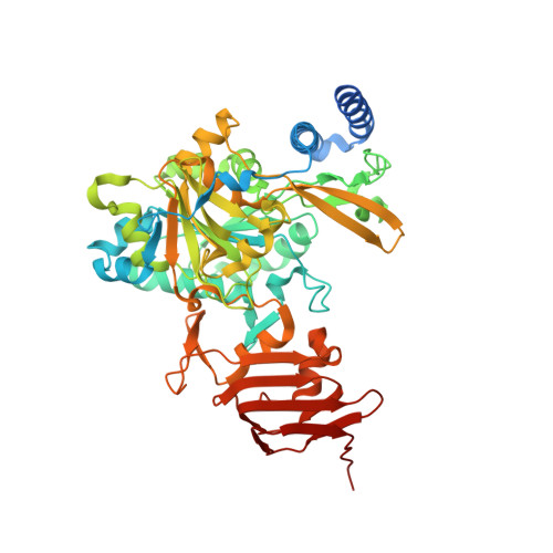
    4R7P

  • PubMed Abstract: 

    In mammals, UDP-glucose pyrophosphorylase (UGP) is the only enzyme capable of activating glucose-1-phosphate (Glc-1-P) to UDP-glucose (UDP-Glc), a metabolite located at the intersection of virtually all metabolic pathways in the mammalian cell. Despite the essential role of its product, the molecular basis of UGP function is poorly understood. Here we report the crystal structure of human UGP in complex with its product UDP-Glc. Beyond providing first insight into the active site architecture, we describe the substrate binding mode and intermolecular interactions in the octameric enzyme that are crucial to its activity. Importantly, the quaternary mechanism identified for human UGP in this study may be common for oligomeric sugar-activating nucleotidyltransferases. Elucidating such mechanisms is essential for understanding nucleotide sugar metabolism and opens the perspective for the development of drugs that specifically inhibit simpler organized nucleotidyltransferases in pathogens.


  • Organizational Affiliation
    • Institute for Cellular Chemistry, Hannover Medical School, Carl-Neuberg-Str. 1, 30625 Hannover, Germany.

Macromolecule Content 

  • Total Structure Weight: 239.45 kDa 
  • Atom Count: 15,591 
  • Modeled Residue Count: 1,935 
  • Deposited Residue Count: 2,104 
  • Unique protein chains: 1

Macromolecules

Find similar proteins by:|  3D Structure
Entity ID: 1
MoleculeChains  Sequence LengthOrganismDetailsImage
UTP--glucose-1-phosphate uridylyltransferase
A, B, C, D
526Homo sapiensMutation(s): 0 
Gene Names: UGP2UGP1
EC: 2.7.7.9
UniProt & NIH Common Fund Data Resources
Find proteins for Q16851 (Homo sapiens)
Explore Q16851 
Go to UniProtKB:  Q16851
PHAROS:  Q16851
GTEx:  ENSG00000169764 
Entity Groups
Sequence Clusters30% Identity50% Identity70% Identity90% Identity95% Identity100% Identity
UniProt GroupQ16851
Sequence Annotations
Expand
Reference Sequence

Small Molecules

Ligands 5 Unique
IDChains Name / Formula / InChI Key2D Diagram3D Interactions
UPG

Query on UPG


Download:Ideal Coordinates CCD File
E [auth A]URIDINE-5'-DIPHOSPHATE-GLUCOSE
C15 H24 N2 O17 P2
HSCJRCZFDFQWRP-JZMIEXBBSA-N
SO4

Query on SO4


Download:Ideal Coordinates CCD File
DA [auth C]
EA [auth C]
F [auth A]
FA [auth C]
G [auth A]
DA [auth C],
EA [auth C],
F [auth A],
FA [auth C],
G [auth A],
H [auth A],
I [auth A],
J [auth A],
K [auth A],
L [auth A],
MA [auth D],
NA [auth D],
OA [auth D],
T [auth B],
U [auth B]
SULFATE ION
O4 S
QAOWNCQODCNURD-UHFFFAOYSA-L
ZN

Query on ZN


Download:Ideal Coordinates CCD File
AA [auth B]
BA [auth B]
CA [auth B]
IA [auth C]
JA [auth C]
AA [auth B],
BA [auth B],
CA [auth B],
IA [auth C],
JA [auth C],
KA [auth C],
LA [auth C],
R [auth A],
S [auth A],
SA [auth D],
TA [auth D]
ZINC ION
Zn
PTFCDOFLOPIGGS-UHFFFAOYSA-N
EDO

Query on EDO


Download:Ideal Coordinates CCD File
HA [auth C]
N [auth A]
O [auth A]
P [auth A]
Q [auth A]
HA [auth C],
N [auth A],
O [auth A],
P [auth A],
Q [auth A],
RA [auth D],
V [auth B],
W [auth B],
X [auth B],
Y [auth B],
Z [auth B]
1,2-ETHANEDIOL
C2 H6 O2
LYCAIKOWRPUZTN-UHFFFAOYSA-N
ACT

Query on ACT


Download:Ideal Coordinates CCD File
GA [auth C],
M [auth A],
PA [auth D],
QA [auth D]
ACETATE ION
C2 H3 O2
QTBSBXVTEAMEQO-UHFFFAOYSA-M

Experimental Data & Validation

Experimental Data

  • Method: X-RAY DIFFRACTION
  • Resolution: 3.35 Å
  • R-Value Free:  0.247 (Depositor), 0.251 (DCC) 
  • R-Value Work:  0.199 (Depositor), 0.205 (DCC) 
  • R-Value Observed: 0.202 (Depositor) 
Space Group: P 31 2 1
Unit Cell:
Length ( Å )Angle ( ˚ )
a = 138.97α = 90
b = 138.97β = 90
c = 311.62γ = 120
Software Package:
Software NamePurpose
MxCuBEdata collection
AMoREphasing
PHENIXrefinement
XDSdata reduction
SADABSdata scaling

Structure Validation

View Full Validation Report


Ligand Structure Quality Assessment 


Entry History 

Deposition Data

Revision History  (Full details and data files)

  • Version 1.0: 2015-04-22
    Type: Initial release
  • Version 1.1: 2024-02-28
    Changes: Data collection, Database references, Derived calculations