4R2N | pdb_00004r2n

Crystal structure of Rv3772 in complex with its substrate


Experimental Data Snapshot

  • Method: X-RAY DIFFRACTION
  • Resolution: 1.98 Å
  • R-Value Free: 
    0.244 (Depositor), 0.248 (DCC) 
  • R-Value Work: 
    0.208 (Depositor), 0.215 (DCC) 
  • R-Value Observed: 
    0.209 (Depositor) 

Starting Model: experimental
View more details

wwPDB Validation 3D Report Full Report

Validation slider image for 4R2N

Ligand Structure Quality Assessment 


This is version 1.2 of the entry. See complete history

Literature

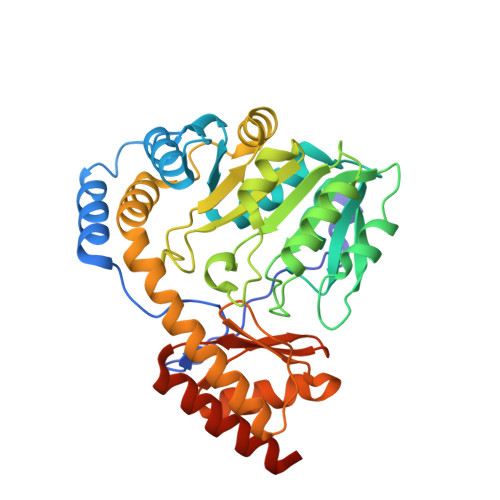
Crystal structures of Mycobacterium tuberculosis HspAT and ArAT reveal structural basis of their distinct substrate specificities

Nasir, N.Anant, A.Vyas, R.Biswal, B.K.

(2016) Sci Rep 6: 18880-18880

  • DOI: https://doi.org/10.1038/srep18880
  • Primary Citation Related Structures: 
    4R2N, 4R5Z

  • PubMed Abstract: 

    Aminotransferases of subfamily Iβ, which include histidinol phosphate aminotransferases (HspATs) and aromatic amino acid aminotransferases (ArATs), are structurally similar but possess distinct substrate specificities. This study, encompassing structural and biochemical characterisation of HspAT and ArAT from Mycobacterium tuberculosis demonstrates that the residues lining the substrate binding pocket and N-terminal lid are the primary determinants of their substrate specificities. In mHspAT, hydrophilic residues in the substrate binding pocket and N-terminal lid allow the entry and binding of its preferential substrate, Hsp. On the other hand, the hydrophobic nature of both the substrate binding pocket and the N-terminal lid of mArAT is responsible for the discrimination of a polar substrate such as Hsp, while facilitating the binding of Phe and other aromatic residues such as Tyr and Trp. In addition, the present study delineates the ligand induced conformational rearrangements, providing insights into the plasticity of aminotransferases. Furthermore, the study also demonstrates that the adventitiously bound ligand 2-(N-morpholino)ethanesulfonic acid (MES) is indeed a specific inhibitor of HspAT. These results suggest that previously untapped morpholine-ring scaffold compounds could be explored for the design of new anti-TB agents.


  • Organizational Affiliation
    • Protein Crystallography Laboratory, National Institute of Immunology, Aruna Asaf Ali Marg, New Delhi, Delhi, 110067, India.

Macromolecule Content 

  • Total Structure Weight: 161.42 kDa 
  • Atom Count: 11,881 
  • Modeled Residue Count: 1,412 
  • Deposited Residue Count: 1,468 
  • Unique protein chains: 1

Macromolecules

Find similar proteins by:|  3D Structure
Entity ID: 1
MoleculeChains  Sequence LengthOrganismDetailsImage
Putative phenylalanine aminotransferase
A, B, C, D
367Mycobacterium tuberculosis H37RvMutation(s): 0 
Gene Names: patP425_03929RVBD_3772
EC: 2.6.1 (PDB Primary Data), 2.6.1.57 (UniProt)
UniProt
Find proteins for P9WML5 (Mycobacterium tuberculosis (strain ATCC 25618 / H37Rv))
Explore P9WML5 
Go to UniProtKB:  P9WML5
Entity Groups
Sequence Clusters30% Identity50% Identity70% Identity90% Identity95% Identity100% Identity
UniProt GroupP9WML5
Sequence Annotations
Expand
Reference Sequence

Small Molecules

Ligands 3 Unique
IDChains Name / Formula / InChI Key2D Diagram3D Interactions
PLP

Query on PLP


Download:Ideal Coordinates CCD File
E [auth A],
H [auth B],
K [auth C],
N [auth D]
PYRIDOXAL-5'-PHOSPHATE
C8 H10 N O6 P
NGVDGCNFYWLIFO-UHFFFAOYSA-N
EPE

Query on EPE


Download:Ideal Coordinates CCD File
G [auth A],
J [auth B],
M [auth C],
P [auth D]
4-(2-HYDROXYETHYL)-1-PIPERAZINE ETHANESULFONIC ACID
C8 H18 N2 O4 S
JKMHFZQWWAIEOD-UHFFFAOYSA-N
PHE

Query on PHE


Download:Ideal Coordinates CCD File
F [auth A],
I [auth B],
L [auth C],
O [auth D]
PHENYLALANINE
C9 H11 N O2
COLNVLDHVKWLRT-QMMMGPOBSA-N

Experimental Data & Validation

Experimental Data

  • Method: X-RAY DIFFRACTION
  • Resolution: 1.98 Å
  • R-Value Free:  0.244 (Depositor), 0.248 (DCC) 
  • R-Value Work:  0.208 (Depositor), 0.215 (DCC) 
  • R-Value Observed: 0.209 (Depositor) 
Space Group: P 21 21 21
Unit Cell:
Length ( Å )Angle ( ˚ )
a = 55.414α = 90
b = 164.46β = 90
c = 178.515γ = 90
Software Package:
Software NamePurpose
MxCuBEdata collection
PHASERphasing
REFMACrefinement
HKL-2000data reduction
SCALEPACKdata scaling

Structure Validation

View Full Validation Report


Ligand Structure Quality Assessment 


Entry History 

Deposition Data

Revision History  (Full details and data files)

  • Version 1.0: 2015-08-12
    Type: Initial release
  • Version 1.1: 2016-12-07
    Changes: Database references
  • Version 1.2: 2023-11-08
    Changes: Data collection, Database references, Derived calculations, Refinement description