


Crystal Structure of Thermophilic L-Arabinose Isomerase with L-Arabitol from Geobacillus kaustophilus
Choi, J.M., Lee, Y.J., Lee, D.W., Lee, S.H.To be published.
Experimental Data Snapshot
Starting Model: experimental
View more details
Entity ID: 1 | |||||
|---|---|---|---|---|---|
| Molecule | Chains | Sequence Length | Organism | Details | Image |
| L-arabinose isomerase | 497 | Geobacillus kaustophilus HTA426 | Mutation(s): 0 Gene Names: araA, GK1904 EC: 5.3.1.4 | 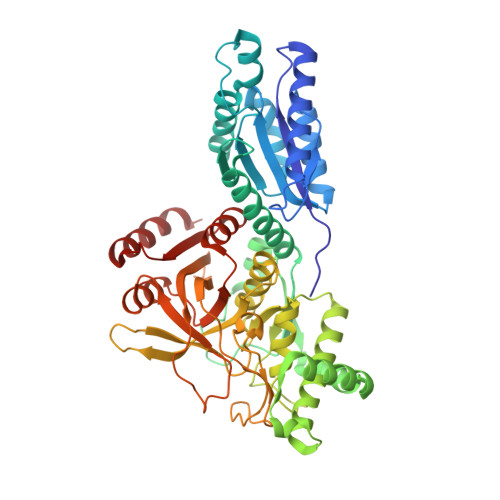 | |
UniProt | |||||
Entity Groups | |||||
| Sequence Clusters | 30% Identity50% Identity70% Identity90% Identity95% Identity100% Identity | ||||
| UniProt Group | Q5KYP7 | ||||
Sequence AnnotationsExpand | |||||
Reference Sequence | |||||
| Ligands 2 Unique | |||||
|---|---|---|---|---|---|
| ID | Chains | Name / Formula / InChI Key | 2D Diagram | 3D Interactions | |
| SST Download:Ideal Coordinates CCD File | G [auth A] H [auth A] J [auth B] L [auth C] M [auth C] | L-arabinitol C5 H12 O5 HEBKCHPVOIAQTA-IMJSIDKUSA-N |  | ||
| MN Download:Ideal Coordinates CCD File | I [auth A] K [auth B] N [auth C] O [auth D] Q [auth E] | MANGANESE (II) ION Mn WAEMQWOKJMHJLA-UHFFFAOYSA-N |  | ||
| Length ( Å ) | Angle ( ˚ ) |
|---|---|
| a = 118.514 | α = 90 |
| b = 146.263 | β = 90 |
| c = 215.677 | γ = 90 |
| Software Name | Purpose |
|---|---|
| HKL-2000 | data collection |
| MOLREP | phasing |
| PHENIX | refinement |
| HKL-2000 | data reduction |
| HKL-2000 | data scaling |


