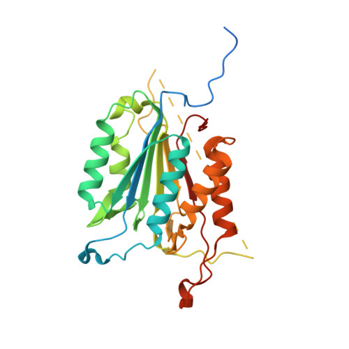
4QTX

Caspase-3 Y195A


Experimental Data Snapshot

  • Method: X-RAY DIFFRACTION
  • Resolution: 1.97 Å
  • R-Value Free: 0.259 
  • R-Value Work: 0.199 
  • R-Value Observed: 0.205 

wwPDB Validation   3D Report Full Report


This is version 1.3 of the entry. See complete history


Literature

Modifying caspase-3 activity by altering allosteric networks.

Cade, C.Swartz, P.MacKenzie, S.H.Clark, A.C.

(2014) Biochemistry 53: 7582-7595

  • DOI: https://doi.org/10.1021/bi500874k
  • Primary Citation of Related Structures:  
    4QTX, 4QTY, 4QU0, 4QU5, 4QU8, 4QU9, 4QUA, 4QUB, 4QUD, 4QUE, 4QUG, 4QUH, 4QUI, 4QUJ, 4QUL

  • PubMed Abstract: 

    Caspases have several allosteric sites that bind small molecules or peptides. Allosteric regulators are known to affect caspase enzyme activity, in general, by facilitating large conformational changes that convert the active enzyme to a zymogen-like form in which the substrate-binding pocket is disordered. Mutations in presumed allosteric networks also decrease activity, although large structural changes are not observed. Mutation of the central V266 to histidine in the dimer interface of caspase-3 inactivates the enzyme by introducing steric clashes that may ultimately affect positioning of a helix on the protein surface. The helix is thought to connect several residues in the active site to the allosteric dimer interface. In contrast to the effects of small molecule allosteric regulators, the substrate-binding pocket is intact in the mutant, yet the enzyme is inactive. We have examined the putative allosteric network, in particular the role of helix 3, by mutating several residues in the network. We relieved steric clashes in the context of caspase-3(V266H), and we show that activity is restored, particularly when the restorative mutation is close to H266. We also mimicked the V266H mutant by introducing steric clashes elsewhere in the allosteric network, generating several mutants with reduced activity. Overall, the data show that the caspase-3 native ensemble includes the canonical active state as well as an inactive conformation characterized by an intact substrate-binding pocket, but with an altered helix 3. The enzyme activity reflects the relative population of each species in the native ensemble.


  • Organizational Affiliation

    Department of Molecular and Structural Biochemistry and ‡Center for Comparative Medicine and Translational Research, North Carolina State University , Raleigh, North Carolina 27695, United States.


Macromolecules
Find similar proteins by:  (by identity cutoff)  |  3D Structure
Entity ID: 1
MoleculeChains Sequence LengthOrganismDetailsImage
Caspase-3277Homo sapiensMutation(s): 1 
Gene Names: CASP3CPP32
EC: 3.4.22.56
UniProt & NIH Common Fund Data Resources
Find proteins for P42574 (Homo sapiens)
Explore P42574 
Go to UniProtKB:  P42574
PHAROS:  P42574
GTEx:  ENSG00000164305 
Entity Groups  
Sequence Clusters30% Identity50% Identity70% Identity90% Identity95% Identity100% Identity
UniProt GroupP42574
Sequence Annotations
Expand
  • Reference Sequence

Find similar proteins by:  Sequence   |   3D Structure  

Entity ID: 2
MoleculeChains Sequence LengthOrganismDetailsImage
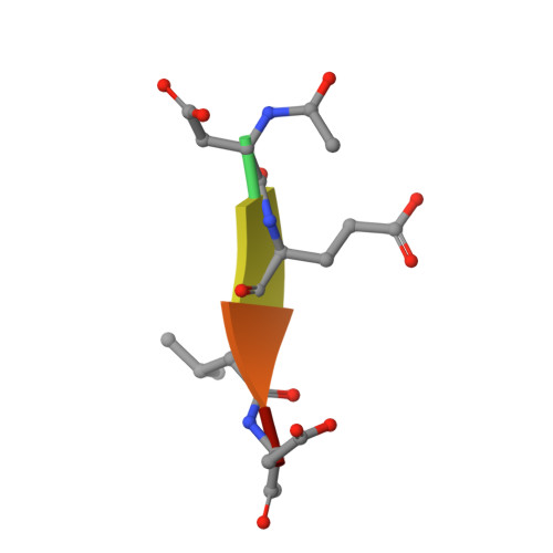
ACE-ASP-GLU-VAL-ASP-CHLOROMETHYLKETONE INHIBITORB [auth E]6N/AMutation(s): 0 
Sequence Annotations
Expand
  • Reference Sequence
Small Molecules
Ligands 1 Unique
IDChains Name / Formula / InChI Key2D Diagram3D Interactions
ACT
Query on ACT

Download Ideal Coordinates CCD File 
C [auth A]ACETATE ION
C2 H3 O2
QTBSBXVTEAMEQO-UHFFFAOYSA-M
Biologically Interesting Molecules (External Reference) 1 Unique
Experimental Data & Validation

Experimental Data

  • Method: X-RAY DIFFRACTION
  • Resolution: 1.97 Å
  • R-Value Free: 0.259 
  • R-Value Work: 0.199 
  • R-Value Observed: 0.205 
  • Space Group: I 2 2 2
Unit Cell:
Length ( Å )Angle ( ˚ )
a = 68.644α = 90
b = 84.493β = 90
c = 96.487γ = 90
Software Package:
Software NamePurpose
MAR345data collection
SERGUIdata collection
PHENIXmodel building
PHENIXrefinement
DENZOdata reduction
SCALEPACKdata scaling
PHENIXphasing

Structure Validation

View Full Validation Report


Entry History 

Deposition Data

Revision History  (Full details and data files)

  • Version 1.0: 2014-11-05
    Type: Initial release
  • Version 1.1: 2014-12-24
    Changes: Database references
  • Version 1.2: 2017-11-22
    Changes: Refinement description
  • Version 1.3: 2024-11-20
    Changes: Data collection, Database references, Derived calculations, Structure summary