4QPM | pdb_00004qpm

Structure of Bub1 kinase domain


Experimental Data Snapshot

  • Method: X-RAY DIFFRACTION
  • Resolution: 2.20 Å
  • R-Value Free: 
    0.269 (Depositor), 0.273 (DCC) 
  • R-Value Work: 
    0.229 (Depositor), 0.235 (DCC) 
  • R-Value Observed: 
    0.231 (Depositor) 

wwPDB Validation   3D Report Full Report


Ligand Structure Quality Assessment 

Created with Raphaël 2.3.0Worse 01 BetterLigand structure goodness of fit to experimental dataBest fitted ADPClick on this verticalbar to view details

This is version 1.2 of the entry. See complete history


Literature

Substrate-Specific Activation of the Mitotic Kinase Bub1 through Intramolecular Autophosphorylation and Kinetochore Targeting.

Lin, Z.Jia, L.Tomchick, D.R.Luo, X.Yu, H.

(2014) Structure 22: 1616-1627

  • DOI: https://doi.org/10.1016/j.str.2014.08.020
  • Primary Citation of Related Structures:  
    4QPM

  • PubMed Abstract: 

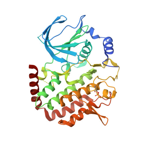
    During mitosis of human cells, the kinase Bub1 orchestrates chromosome segregation through phosphorylating histone H2A and the anaphase-promoting complex/cyclosome activator Cdc20. Bub1-mediated H2A-T120 phosphorylation (H2A-pT120) at kinetochores promotes centromeric sister-chromatid cohesion, whereas Cdc20 phosphorylation by Bub1 contributes to spindle checkpoint signaling. Here, we show that phosphorylation at the P+1 substrate-binding loop of human Bub1 enhances its activity toward H2A but has no effect on its activity toward Cdc20. We determine the crystal structure of phosphorylated Bub1. A comparison between structures of phosphorylated and unphosphorylated Bub1 reveals phosphorylation-triggered reorganization of the P+1 loop. This activating phosphorylation of Bub1 is constitutive during the cell cycle. Enrichment of H2A-pT120 at mitotic kinetochores requires kinetochore targeting of Bub1. The P+1 loop phosphorylation of Bub1 appears to occur through intramolecular autophosphorylation. Our study provides structural and functional insights into substrate-specific regulation of a key mitotic kinase and expands the repertoire of kinase activation mechanisms.


  • Organizational Affiliation

    Howard Hughes Medical Institute, University of Texas Southwestern Medical Center, 6001 Forest Park Road, Dallas, TX 75390, USA; Department of Pharmacology, University of Texas Southwestern Medical Center, 6001 Forest Park Road, Dallas, TX 75390, USA.


Macromolecules
Find similar proteins by:  (by identity cutoff)  |  3D Structure
Entity ID: 1
MoleculeChains Sequence LengthOrganismDetailsImage
Mitotic checkpoint serine/threonine-protein kinase BUB1
A, B
356Homo sapiensMutation(s): 0 
Gene Names: BUB1BUB1L
EC: 2.7.11.1
UniProt & NIH Common Fund Data Resources
Find proteins for O43683 (Homo sapiens)
Explore O43683 
Go to UniProtKB:  O43683
PHAROS:  O43683
GTEx:  ENSG00000169679 
Entity Groups  
Sequence Clusters30% Identity50% Identity70% Identity90% Identity95% Identity100% Identity
UniProt GroupO43683
Sequence Annotations
Expand
  • Reference Sequence
Small Molecules
Ligands 3 Unique
IDChains Name / Formula / InChI Key2D Diagram3D Interactions
ADP
Query on ADP

Download Ideal Coordinates CCD File 
C [auth A],
G [auth B]
ADENOSINE-5'-DIPHOSPHATE
C10 H15 N5 O10 P2
XTWYTFMLZFPYCI-KQYNXXCUSA-N
CL
Query on CL

Download Ideal Coordinates CCD File 
F [auth A],
J [auth B],
K [auth B]
CHLORIDE ION
Cl
VEXZGXHMUGYJMC-UHFFFAOYSA-M
MG
Query on MG

Download Ideal Coordinates CCD File 
D [auth A],
E [auth A],
H [auth B],
I [auth B]
MAGNESIUM ION
Mg
JLVVSXFLKOJNIY-UHFFFAOYSA-N
Modified Residues  1 Unique
IDChains TypeFormula2D DiagramParent
SEP
Query on SEP
A, B
L-PEPTIDE LINKINGC3 H8 N O6 PSER
Experimental Data & Validation

Experimental Data

  • Method: X-RAY DIFFRACTION
  • Resolution: 2.20 Å
  • R-Value Free:  0.269 (Depositor), 0.273 (DCC) 
  • R-Value Work:  0.229 (Depositor), 0.235 (DCC) 
  • R-Value Observed: 0.231 (Depositor) 
Space Group: P 1 21 1
Unit Cell:
Length ( Å )Angle ( ˚ )
a = 90.013α = 90
b = 47.18β = 107.4
c = 93.146γ = 90
Software Package:
Software NamePurpose
HKL-3000data collection
PHASESphasing
PHENIXrefinement
HKL-3000data reduction
HKL-3000data scaling

Structure Validation

View Full Validation Report


Ligand Structure Quality Assessment 

Created with Raphaël 2.3.0Worse 01 BetterLigand structure goodness of fit to experimental dataBest fitted ADPClick on this verticalbar to view details

Entry History 

Deposition Data

Revision History  (Full details and data files)

  • Version 1.0: 2014-10-22
    Type: Initial release
  • Version 1.1: 2014-12-24
    Changes: Database references
  • Version 1.2: 2024-11-06
    Changes: Data collection, Database references, Derived calculations, Structure summary