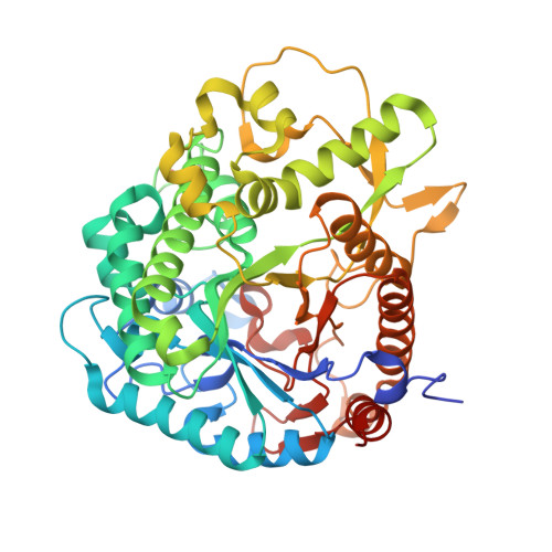
4QLJ

Crystal structure of rice BGlu1 E386G/Y341A/Q187A mutant complexed with cellotetraose


Experimental Data Snapshot

  • Method: X-RAY DIFFRACTION
  • Resolution: 1.95 Å
  • R-Value Free: 0.202 
  • R-Value Work: 0.173 
  • R-Value Observed: 0.175 

Starting Model: experimental
View more details

wwPDB Validation   3D Report Full Report


This is version 2.1 of the entry. See complete history


Literature

Effects of active site cleft residues on oligosaccharide binding, hydrolysis, and glycosynthase activities of rice BGlu1 and its mutants

Pengthaisong, S.Ketudat Cairns, J.R.

(2014) Protein Sci 23: 1738-1752

  • DOI: https://doi.org/10.1002/pro.2556
  • Primary Citation of Related Structures:  
    4QLJ, 4QLK, 4QLL

  • PubMed Abstract: 

    Rice BGlu1 (Os3BGlu7) is a glycoside hydrolase family 1 β-glucosidase that hydrolyzes cellooligosaccharides with increasing efficiency as the degree of polymerization (DP) increases from 2 to 6, indicating six subsites for glucosyl residue binding. Five subsites have been identified in X-ray crystal structures of cellooligosaccharide complexes with its E176Q acid-base and E386G nucleophile mutants. X-ray crystal structures indicate that cellotetraose binds in a similar mode in BGlu1 E176Q and E386G, but in a different mode in the BGlu1 E386G/Y341A variant, in which glucosyl residue 4 (Glc4) interacts with Q187 instead of the eliminated phenolic group of Y341. Here, we found that the Q187A mutation has little effect on BGlu1 cellooligosaccharide hydrolysis activity or oligosaccharide binding in BGlu1 E176Q, and only slight effects on BGlu1 E386G glycosynthase activity. X-ray crystal structures showed that cellotetraose binds in a different position in BGlu1 E176Q/Y341A, in which it interacts directly with R178 and W337, and the Q187A mutation had little effect on cellotetraose binding. Mutations of R178 and W337 to A had significant and nonadditive effects on oligosaccharide hydrolysis by BGlu1, pNPGlc cleavage and cellooligosaccharide inhibition of BGlu1 E176Q and BGlu1 E386G glycosynthase activity. Hydrolysis activity was partially rescued by Y341 for longer substrates, suggesting stacking of Glc4 on Y341 stabilizes binding of cellooligosaccharides in the optimal position for hydrolysis. This analysis indicates that complex interactions between active site cleft residues modulate substrate binding and hydrolysis.


  • Organizational Affiliation

    School of Biochemistry, Institute of Science, and Center for Biomolecular Structure, Function and Application, Suranaree University of Technology, Nakhon Ratchasima, 30000, Thailand.


Macromolecules
Find similar proteins by:  (by identity cutoff)  |  3D Structure
Entity ID: 1
MoleculeChains Sequence LengthOrganismDetailsImage
Beta-glucosidase 7
A, B
481Oryza sativa Japonica GroupMutation(s): 3 
Gene Names: BGLU1BGLU7LOC_Os03g49600Os03g0703000Os3BGlu7OSJNBa0004L11.16
EC: 3.2.1.21
UniProt
Find proteins for Q75I93 (Oryza sativa subsp. japonica)
Explore Q75I93 
Go to UniProtKB:  Q75I93
Entity Groups  
Sequence Clusters30% Identity50% Identity70% Identity90% Identity95% Identity100% Identity
UniProt GroupQ75I93
Sequence Annotations
Expand
  • Reference Sequence
Oligosaccharides

Help

Entity ID: 2
MoleculeChains Length2D Diagram Glycosylation3D Interactions
beta-D-glucopyranose-(1-4)-beta-D-glucopyranose-(1-4)-beta-D-glucopyranose-(1-4)-beta-D-glucopyranose
C, D, E
4N/A
Glycosylation Resources
GlyTouCan:  G00025MO
GlyCosmos:  G00025MO
GlyGen:  G00025MO
Biologically Interesting Molecules (External Reference) 1 Unique
Experimental Data & Validation

Experimental Data

  • Method: X-RAY DIFFRACTION
  • Resolution: 1.95 Å
  • R-Value Free: 0.202 
  • R-Value Work: 0.173 
  • R-Value Observed: 0.175 
  • Space Group: P 21 21 21
Unit Cell:
Length ( Å )Angle ( ˚ )
a = 79.505α = 90
b = 101.239β = 90
c = 127.349γ = 90
Software Package:
Software NamePurpose
HKL-2000data collection
Cootmodel building
REFMACrefinement
HKL-2000data reduction
HKL-2000data scaling

Structure Validation

View Full Validation Report


Entry History 

Deposition Data

Revision History  (Full details and data files)

  • Version 1.0: 2015-06-17
    Type: Initial release
  • Version 1.1: 2015-06-24
    Changes: Other
  • Version 2.0: 2020-07-29
    Type: Remediation
    Reason: Carbohydrate remediation
    Changes: Atomic model, Data collection, Database references, Derived calculations, Non-polymer description, Structure summary
  • Version 2.1: 2023-11-08
    Changes: Data collection, Database references, Refinement description, Structure summary