4QL0 | pdb_00004ql0

Crystal Structure Analysis of the Membrane Transporter FhaC (double mutant V169T, I176N)


Experimental Data Snapshot

  • Method: X-RAY DIFFRACTION
  • Resolution: 2.50 Å
  • R-Value Free: 
    0.259 (Depositor), 0.268 (DCC) 
  • R-Value Work: 
    0.217 (Depositor), 0.223 (DCC) 
  • R-Value Observed: 
    0.221 (Depositor) 

Starting Model: experimental
View more details

wwPDB Validation 3D Report Full Report

Validation slider image for 4QL0

Ligand Structure Quality Assessment 


This is version 1.4 of the entry. See complete history

Literature

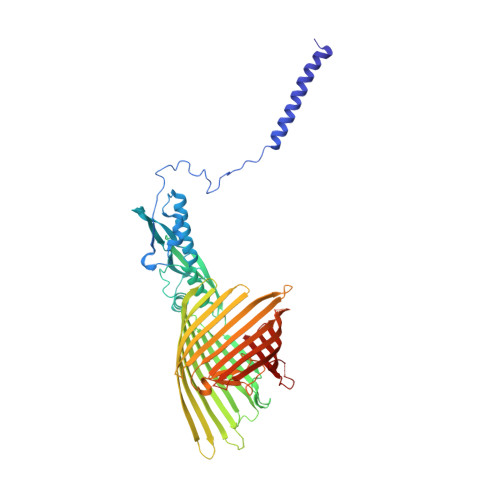
Conserved Omp85 lid-lock structure and substrate recognition in FhaC

Maier, T.Clantin, B.Gruss, F.Dewitte, F.Delattre, A.S.Jacob-Dubuisson, F.Hiller, S.Villeret, V.

(2015) Nat Commun 6: 7452-7452

  • DOI: https://doi.org/10.1038/ncomms8452
  • Primary Citation Related Structures: 
    4QKY, 4QL0

  • PubMed Abstract: 

    Omp85 proteins mediate translocation of polypeptide substrates across and into cellular membranes. They share a common architecture comprising substrate-interacting POTRA domains, a C-terminal 16-stranded β-barrel pore and two signature motifs located on the inner barrel wall and at the tip of the extended L6 loop. The observation of two distinct conformations of the L6 loop in the available Omp85 structures previously suggested a functional role of conformational changes in L6 in the Omp85 mechanism. Here we present a 2.5 Å resolution structure of a variant of the Omp85 secretion protein FhaC, in which the two signature motifs interact tightly and form the conserved 'lid lock'. Reanalysis of previous structural data shows that L6 adopts the same, conserved resting state position in all available Omp85 structures. The FhaC variant structure further reveals a competitive mechanism for the regulation of substrate binding mediated by the linker to the N-terminal plug helix H1.


  • Organizational Affiliation
    • Biozentrum, University of Basel, Klingelbergstr. 70, Basel 4056, Switzerland.

Macromolecule Content 

  • Total Structure Weight: 63.36 kDa 
  • Atom Count: 4,300 
  • Modeled Residue Count: 520 
  • Deposited Residue Count: 554 
  • Unique protein chains: 1

Macromolecules

Find similar proteins by:|  3D Structure
Entity ID: 1
MoleculeChains  Sequence LengthOrganismDetailsImage
Filamentous hemagglutinin transporter protein FhaC554Bordetella pertussis Tohama IMutation(s): 2 
Gene Names: BP1884fhaC
Membrane Entity: Yes 
UniProt
Find proteins for P35077 (Bordetella pertussis (strain Tohama I / ATCC BAA-589 / NCTC 13251))
Explore P35077 
Go to UniProtKB:  P35077
Entity Groups
Sequence Clusters30% Identity50% Identity70% Identity90% Identity95% Identity100% Identity
UniProt GroupP35077
Sequence Annotations
Expand
Reference Sequence

Small Molecules

Ligands 7 Unique
IDChains Name / Formula / InChI Key2D Diagram3D Interactions
BOG

Query on BOG


Download:Ideal Coordinates CCD File
D [auth A],
E [auth A],
G [auth A]
octyl beta-D-glucopyranoside
C14 H28 O6
HEGSGKPQLMEBJL-RKQHYHRCSA-N
P6G

Query on P6G


Download:Ideal Coordinates CCD File
J [auth A]HEXAETHYLENE GLYCOL
C12 H26 O7
IIRDTKBZINWQAW-UHFFFAOYSA-N
1PE

Query on 1PE


Download:Ideal Coordinates CCD File
H [auth A]PENTAETHYLENE GLYCOL
C10 H22 O6
JLFNLZLINWHATN-UHFFFAOYSA-N
PG4

Query on PG4


Download:Ideal Coordinates CCD File
I [auth A]TETRAETHYLENE GLYCOL
C8 H18 O5
UWHCKJMYHZGTIT-UHFFFAOYSA-N
PGE

Query on PGE


Download:Ideal Coordinates CCD File
B [auth A]TRIETHYLENE GLYCOL
C6 H14 O4
ZIBGPFATKBEMQZ-UHFFFAOYSA-N
PEG

Query on PEG


Download:Ideal Coordinates CCD File
C [auth A]DI(HYDROXYETHYL)ETHER
C4 H10 O3
MTHSVFCYNBDYFN-UHFFFAOYSA-N
PO4

Query on PO4


Download:Ideal Coordinates CCD File
F [auth A]PHOSPHATE ION
O4 P
NBIIXXVUZAFLBC-UHFFFAOYSA-K

Experimental Data & Validation

Experimental Data

  • Method: X-RAY DIFFRACTION
  • Resolution: 2.50 Å
  • R-Value Free:  0.259 (Depositor), 0.268 (DCC) 
  • R-Value Work:  0.217 (Depositor), 0.223 (DCC) 
  • R-Value Observed: 0.221 (Depositor) 
Space Group: C 2 2 21
Unit Cell:
Length ( Å )Angle ( ˚ )
a = 106.38α = 90
b = 136.95β = 90
c = 110.97γ = 90
Software Package:
Software NamePurpose
HKL-2000data collection
BUSTERrefinement
XDSdata reduction
XDSdata scaling

Structure Validation

View Full Validation Report


Ligand Structure Quality Assessment 


Entry History 

Deposition Data

Revision History  (Full details and data files)

  • Version 1.0: 2015-06-17
    Type: Initial release
  • Version 1.1: 2015-06-24
    Changes: Derived calculations
  • Version 1.2: 2016-12-14
    Changes: Database references
  • Version 1.3: 2020-07-29
    Type: Remediation
    Reason: Carbohydrate remediation
    Changes: Data collection, Database references, Derived calculations, Structure summary
  • Version 1.4: 2023-11-08
    Changes: Data collection, Database references, Refinement description, Structure summary