4QGO

Crystal structure of NucA from Streptococcus agalactiae with no metal bound


Experimental Data Snapshot

  • Method: X-RAY DIFFRACTION
  • Resolution: 1.50 Å
  • R-Value Free: 0.180 
  • R-Value Work: 0.153 
  • R-Value Observed: 0.154 

wwPDB Validation   3D Report Full Report


This is version 1.1 of the entry. See complete history


Literature

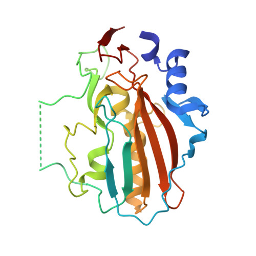
Structural characterization of the virulence factor nuclease A from Streptococcus agalactiae.

Moon, A.F.Gaudu, P.Pedersen, L.C.

(2014) Acta Crystallogr D Biol Crystallogr 70: 2937-2949

  • DOI: https://doi.org/10.1107/S1399004714019725
  • Primary Citation of Related Structures:  
    4QGO, 4QH0

  • PubMed Abstract: 

    The group B pathogen Streptococcus agalactiae commonly populates the human gut and urogenital tract, and is a major cause of infection-based mortality in neonatal infants and in elderly or immunocompromised adults. Nuclease A (GBS_NucA), a secreted DNA/RNA nuclease, serves as a virulence factor for S. agalactiae, facilitating bacterial evasion of the human innate immune response. GBS_NucA efficiently degrades the DNA matrix component of neutrophil extracellular traps (NETs), which attempt to kill and clear invading bacteria during the early stages of infection. In order to better understand the mechanisms of DNA substrate binding and catalysis of GBS_NucA, the high-resolution structure of a catalytically inactive mutant (H148G) was solved by X-ray crystallography. Several mutants on the surface of GBS_NucA which might influence DNA substrate binding and catalysis were generated and evaluated using an imidazole chemical rescue technique. While several of these mutants severely inhibited nuclease activity, two mutants (K146R and Q183A) exhibited significantly increased activity. These structural and biochemical studies have greatly increased our understanding of the mechanism of action of GBS_NucA in bacterial virulence and may serve as a foundation for the structure-based drug design of antibacterial compounds targeted to S. agalactiae.


  • Organizational Affiliation

    Laboratory of Structural Biology, National Institute of Environmental Health Sciences, National Institutes of Health, Research Triangle Park, NC 27709, USA.


Macromolecules
Find similar proteins by:  (by identity cutoff)  |  3D Structure
Entity ID: 1
MoleculeChains Sequence LengthOrganismDetailsImage
DNA-entry nuclease (Competence-specific nuclease)
A, B
225Streptococcus agalactiae ILRI112Mutation(s): 1 
Gene Names: SAIL_8320
Entity Groups  
Sequence Clusters30% Identity50% Identity70% Identity90% Identity95% Identity100% Identity
Sequence Annotations
Expand
  • Reference Sequence
Small Molecules
Ligands 3 Unique
IDChains Name / Formula / InChI Key2D Diagram3D Interactions
SO4
Query on SO4

Download Ideal Coordinates CCD File 
C [auth A]
D [auth A]
E [auth A]
F [auth A]
G [auth A]
C [auth A],
D [auth A],
E [auth A],
F [auth A],
G [auth A],
K [auth B],
L [auth B],
M [auth B],
N [auth B]
SULFATE ION
O4 S
QAOWNCQODCNURD-UHFFFAOYSA-L
EDO
Query on EDO

Download Ideal Coordinates CCD File 
I [auth A],
O [auth B]
1,2-ETHANEDIOL
C2 H6 O2
LYCAIKOWRPUZTN-UHFFFAOYSA-N
CL
Query on CL

Download Ideal Coordinates CCD File 
H [auth A],
J [auth A],
P [auth B],
Q [auth B]
CHLORIDE ION
Cl
VEXZGXHMUGYJMC-UHFFFAOYSA-M
Experimental Data & Validation

Experimental Data

  • Method: X-RAY DIFFRACTION
  • Resolution: 1.50 Å
  • R-Value Free: 0.180 
  • R-Value Work: 0.153 
  • R-Value Observed: 0.154 
  • Space Group: P 1
Unit Cell:
Length ( Å )Angle ( ˚ )
a = 37.591α = 109.4
b = 57.131β = 97.08
c = 62.422γ = 90.16
Software Package:
Software NamePurpose
SERGUIdata collection
PHASERphasing
PHENIXrefinement
HKL-2000data reduction
HKL-2000data scaling

Structure Validation

View Full Validation Report


Entry History 

Deposition Data

Revision History  (Full details and data files)

  • Version 1.0: 2014-11-19
    Type: Initial release
  • Version 1.1: 2023-09-20
    Changes: Data collection, Database references, Derived calculations, Refinement description