4Q3R

Crystal structure of Schistosoma mansoni arginase in complex with inhibitor ABHDP


Experimental Data Snapshot

  • Method: X-RAY DIFFRACTION
  • Resolution: 2.17 Å
  • R-Value Free: 0.206 
  • R-Value Work: 0.174 
  • R-Value Observed: 0.176 

Starting Model: experimental
View more details

wwPDB Validation   3D Report Full Report


Ligand Structure Quality Assessment 


This is version 1.4 of the entry. See complete history


Literature

Crystal Structure of Schistosoma mansoni Arginase, a Potential Drug Target for the Treatment of Schistosomiasis.

Hai, Y.Edwards, J.E.Van Zandt, M.C.Hoffmann, K.F.Christianson, D.W.

(2014) Biochemistry 53: 4671-4684

  • DOI: https://doi.org/10.1021/bi5004519
  • Primary Citation of Related Structures:  
    4Q3P, 4Q3Q, 4Q3R, 4Q3S, 4Q3T, 4Q3U, 4Q3V, 4Q40, 4Q41, 4Q42

  • PubMed Abstract: 

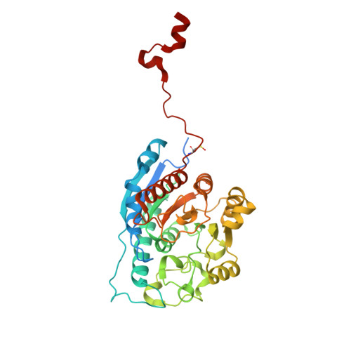
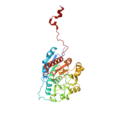
    The X-ray crystal structure of arginase from Schistosoma mansoni (SmARG) and the structures of its complexes with several amino acid inhibitors have been determined at atomic resolution. SmARG is a binuclear manganese metalloenzyme that catalyzes the hydrolysis of l-arginine to form l-ornithine and urea, and this enzyme is upregulated in all forms of the parasite that interact with the human host. Current hypotheses suggest that parasitic arginases could play a role in host immune evasion by depleting pools of substrate l-arginine that would otherwise be utilized for NO biosynthesis and NO-dependent processes in the immune response. Although the amino acid sequence of SmARG is only 42% identical with that of human arginase I, residues important for substrate binding and catalysis are strictly conserved. In general, classical amino acid inhibitors such as 2(S)-amino-6-boronohexanoic acid (ABH) tend to bind more weakly to SmARG than to human arginase I despite identical inhibitor binding modes in each enzyme active site. The identification of a patch on the enzyme surface capable of accommodating the additional Cα substitutent of an α,α-disubstituted amino acid inhibitor suggests that such inhibitors could exhibit higher affinity and biological activity. The structures of SmARG complexed with two different α,α-disubstituted derivatives of ABH are presented and provide a proof of concept for this approach in the enhancement of enzyme-inhibitor affinity.


  • Organizational Affiliation

    Roy and Diana Vagelos Laboratories, Department of Chemistry, University of Pennsylvania , Philadelphia, Pennsylvania 19104-6323, United States.


Macromolecules
Find similar proteins by:  (by identity cutoff)  |  3D Structure
Entity ID: 1
MoleculeChains Sequence LengthOrganismDetailsImage
Arginase
A, B
385Schistosoma mansoniMutation(s): 0 
Gene Names: Smp_059980
EC: 3.5.3.1
UniProt
Find proteins for Q6WVP6 (Schistosoma mansoni)
Explore Q6WVP6 
Go to UniProtKB:  Q6WVP6
Entity Groups  
Sequence Clusters30% Identity50% Identity70% Identity90% Identity95% Identity100% Identity
UniProt GroupQ6WVP6
Sequence Annotations
Expand
  • Reference Sequence
Find similar proteins by:  (by identity cutoff)  |  3D Structure
Entity ID: 2
MoleculeChains Sequence LengthOrganismDetailsImage
Arginase
C, D
385Schistosoma mansoniMutation(s): 0 
Gene Names: Smp_059980
EC: 3.5.3.1
UniProt
Find proteins for Q6WVP6 (Schistosoma mansoni)
Explore Q6WVP6 
Go to UniProtKB:  Q6WVP6
Entity Groups  
Sequence Clusters30% Identity50% Identity70% Identity90% Identity95% Identity100% Identity
UniProt GroupQ6WVP6
Sequence Annotations
Expand
  • Reference Sequence
Small Molecules
Ligands 6 Unique
IDChains Name / Formula / InChI Key2D Diagram3D Interactions
XA2
Query on XA2

Download Ideal Coordinates CCD File 
FA [auth C],
K [auth A],
LA [auth D],
W [auth B]
(R)-2-amino-6-borono-2-(1-(3,4-dichlorobenzyl)piperidin-4-yl)hexanoic acid
C18 H28 B Cl2 N2 O5
LYUGMNLZENMUTE-GOSISDBHSA-N
PGE
Query on PGE

Download Ideal Coordinates CCD File 
N [auth A]TRIETHYLENE GLYCOL
C6 H14 O4
ZIBGPFATKBEMQZ-UHFFFAOYSA-N
CS
Query on CS

Download Ideal Coordinates CCD File 
BA [auth C]
CA [auth C]
EA [auth C]
G [auth A]
H [auth A]
BA [auth C],
CA [auth C],
EA [auth C],
G [auth A],
H [auth A],
I [auth A],
KA [auth D],
Q [auth B],
R [auth B],
S [auth B],
T [auth B]
CESIUM ION
Cs
NCMHKCKGHRPLCM-UHFFFAOYSA-N
GOL
Query on GOL

Download Ideal Coordinates CCD File 
GA [auth C]
HA [auth C]
L [auth A]
M [auth A]
X [auth B]
GA [auth C],
HA [auth C],
L [auth A],
M [auth A],
X [auth B],
Y [auth B]
GLYCEROL
C3 H8 O3
PEDCQBHIVMGVHV-UHFFFAOYSA-N
MN
Query on MN

Download Ideal Coordinates CCD File 
AA [auth C]
E [auth A]
F [auth A]
IA [auth D]
JA [auth D]
AA [auth C],
E [auth A],
F [auth A],
IA [auth D],
JA [auth D],
O [auth B],
P [auth B],
Z [auth C]
MANGANESE (II) ION
Mn
WAEMQWOKJMHJLA-UHFFFAOYSA-N
CL
Query on CL

Download Ideal Coordinates CCD File 
DA [auth C],
J [auth A],
U [auth B],
V [auth B]
CHLORIDE ION
Cl
VEXZGXHMUGYJMC-UHFFFAOYSA-M
Modified Residues  1 Unique
IDChains TypeFormula2D DiagramParent
CSO
Query on CSO
A, B
L-PEPTIDE LINKINGC3 H7 N O3 SCYS
Experimental Data & Validation

Experimental Data

  • Method: X-RAY DIFFRACTION
  • Resolution: 2.17 Å
  • R-Value Free: 0.206 
  • R-Value Work: 0.174 
  • R-Value Observed: 0.176 
  • Space Group: P 21 3
Unit Cell:
Length ( Å )Angle ( ˚ )
a = 177.488α = 90
b = 177.488β = 90
c = 177.488γ = 90
Software Package:
Software NamePurpose
CBASSdata collection
PHASERphasing
PHENIXrefinement
HKL-2000data reduction
HKL-2000data scaling

Structure Validation

View Full Validation Report


Ligand Structure Quality Assessment 


Entry History 

Deposition Data

Revision History  (Full details and data files)

  • Version 1.0: 2014-07-16
    Type: Initial release
  • Version 1.1: 2014-07-23
    Changes: Database references
  • Version 1.2: 2014-08-06
    Changes: Database references
  • Version 1.3: 2023-09-20
    Changes: Data collection, Database references, Derived calculations, Refinement description
  • Version 1.4: 2023-12-06
    Changes: Data collection