4PZ6

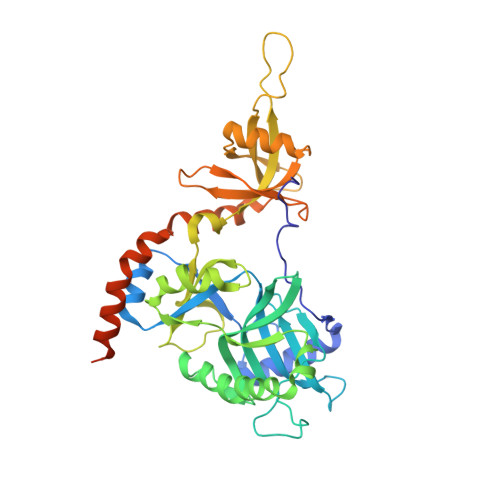
PCE1 guanylyltransferase bound to SER2/SER5 phosphorylated RNA pol II CTD


Experimental Data Snapshot

  • Method: X-RAY DIFFRACTION
  • Resolution: 2.41 Å
  • R-Value Free: 0.233 
  • R-Value Work: 0.180 
  • R-Value Observed: 0.183 

Starting Model: experimental
View more details

wwPDB Validation   3D Report Full Report


Ligand Structure Quality Assessment 


This is version 1.4 of the entry. See complete history


Literature

How an mRNA capping enzyme reads distinct RNA polymerase II and Spt5 CTD phosphorylation codes.

Doamekpor, S.K.Sanchez, A.M.Schwer, B.Shuman, S.Lima, C.D.

(2014) Genes Dev 28: 1323-1336

  • DOI: https://doi.org/10.1101/gad.242768.114
  • Primary Citation of Related Structures:  
    4PZ6, 4PZ7, 4PZ8

  • PubMed Abstract: 

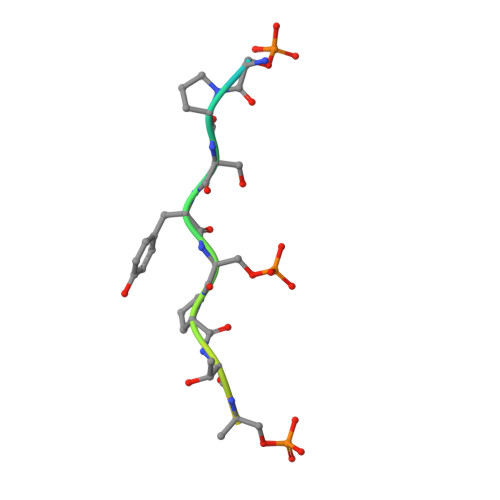
    Interactions between RNA guanylyltransferase (GTase) and the C-terminal domain (CTD) repeats of RNA polymerase II (Pol2) and elongation factor Spt5 are thought to orchestrate cotranscriptional capping of nascent mRNAs. The crystal structure of a fission yeast GTase•Pol2 CTD complex reveals a unique docking site on the nucleotidyl transferase domain for an 8-amino-acid Pol2 CTD segment, S5PPSYSPTS5P, bracketed by two Ser5-PO4 marks. Analysis of GTase mutations that disrupt the Pol2 CTD interface shows that at least one of the two Ser5-PO4-binding sites is required for cell viability and that each site is important for cell growth at 37°C. Fission yeast GTase binds the Spt5 CTD at a separate docking site in the OB-fold domain that captures the Trp4 residue of the Spt5 nonapeptide repeat T(1)PAW(4)NSGSK. A disruptive mutation in the Spt5 CTD-binding site of GTase is synthetically lethal with mutations in the Pol2 CTD-binding site, signifying that the Spt5 and Pol2 CTDs cooperate to recruit capping enzyme in vivo. CTD phosphorylation has opposite effects on the interaction of GTase with Pol2 (Ser5-PO4 is required for binding) versus Spt5 (Thr1-PO4 inhibits binding). We propose that the state of Thr1 phosphorylation comprises a binary "Spt5 CTD code" that is read by capping enzyme independent of and parallel to its response to the state of the Pol2 CTD.


  • Organizational Affiliation

    Structural Biology Program, Sloan-Kettering Institute, New York, New York 10065, USA;


Macromolecules
Find similar proteins by:  (by identity cutoff)  |  3D Structure
Entity ID: 1
MoleculeChains Sequence LengthOrganismDetailsImage
mRNA-capping enzyme subunit alpha
A, B
403Schizosaccharomyces pombe 972h-Mutation(s): 0 
Gene Names: ceg1ceg1/pce1pce1SPBC2F12.08c
EC: 2.7.7.50
UniProt
Find proteins for P40997 (Schizosaccharomyces pombe (strain 972 / ATCC 24843))
Explore P40997 
Go to UniProtKB:  P40997
Entity Groups  
Sequence Clusters30% Identity50% Identity70% Identity90% Identity95% Identity100% Identity
UniProt GroupP40997
Sequence Annotations
Expand
  • Reference Sequence

Find similar proteins by:  Sequence   |   3D Structure  

Entity ID: 2
MoleculeChains Sequence LengthOrganismDetailsImage
DNA-directed RNA polymerase II subunit rpb1C [auth P],
D [auth Q]
21Schizosaccharomyces pombe 972h-Mutation(s): 0 
EC: 2.7.7.6
UniProt
Find proteins for P36594 (Schizosaccharomyces pombe (strain 972 / ATCC 24843))
Explore P36594 
Go to UniProtKB:  P36594
Entity Groups  
Sequence Clusters30% Identity50% Identity70% Identity90% Identity95% Identity100% Identity
UniProt GroupP36594
Sequence Annotations
Expand
  • Reference Sequence
Small Molecules
Ligands 1 Unique
IDChains Name / Formula / InChI Key2D Diagram3D Interactions
GMP
Query on GMP

Download Ideal Coordinates CCD File 
E [auth B]GUANOSINE
C10 H13 N5 O5
NYHBQMYGNKIUIF-UUOKFMHZSA-N
Modified Residues  1 Unique
IDChains TypeFormula2D DiagramParent
SEP
Query on SEP
C [auth P],
D [auth Q]
L-PEPTIDE LINKINGC3 H8 N O6 PSER
Experimental Data & Validation

Experimental Data

  • Method: X-RAY DIFFRACTION
  • Resolution: 2.41 Å
  • R-Value Free: 0.233 
  • R-Value Work: 0.180 
  • R-Value Observed: 0.183 
  • Space Group: P 1 21 1
Unit Cell:
Length ( Å )Angle ( ˚ )
a = 56.406α = 90
b = 78.614β = 91.42
c = 115.716γ = 90
Software Package:
Software NamePurpose
ADSCdata collection
PHASERphasing
PHENIXrefinement
HKL-2000data reduction
SCALEPACKdata scaling

Structure Validation

View Full Validation Report


Ligand Structure Quality Assessment 


Entry History 

Deposition Data

Revision History  (Full details and data files)

  • Version 1.0: 2014-06-25
    Type: Initial release
  • Version 1.1: 2014-07-02
    Changes: Structure summary
  • Version 1.2: 2014-09-24
    Changes: Database references
  • Version 1.3: 2023-09-20
    Changes: Data collection, Database references, Derived calculations, Refinement description
  • Version 1.4: 2024-10-16
    Changes: Structure summary