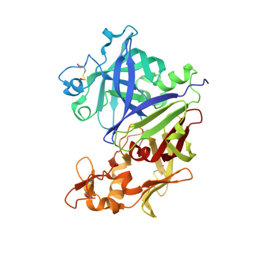
4PYV

Crystal structure of renin in complex with compound4


Experimental Data Snapshot

  • Method: X-RAY DIFFRACTION
  • Resolution: 2.65 Å
  • R-Value Free: 0.241 
  • R-Value Work: 0.201 
  • R-Value Observed: 0.205 

wwPDB Validation   3D Report Full Report


Ligand Structure Quality Assessment 


This is version 1.1 of the entry. See complete history


Literature

Structure-based design of substituted piperidines as a new class of highly efficacious oral direct Renin inhibitors.

Ehara, T.Irie, O.Kosaka, T.Kanazawa, T.Breitenstein, W.Grosche, P.Ostermann, N.Suzuki, M.Kawakami, S.Konishi, K.Hitomi, Y.Toyao, A.Gunji, H.Cumin, F.Schiering, N.Wagner, T.Rigel, D.F.Webb, R.L.Maibaum, J.Yokokawa, F.

(2014) ACS Med Chem Lett 5: 787-792

  • DOI: https://doi.org/10.1021/ml500137b
  • Primary Citation of Related Structures:  
    4PYV, 4Q1N

  • PubMed Abstract: 

    A cis-configured 3,5-disubstituted piperidine direct renin inhibitor, (syn,rac)-1, was discovered as a high-throughput screening hit from a target-family tailored library. Optimization of both the prime and the nonprime site residues flanking the central piperidine transition-state surrogate resulted in analogues with improved potency and pharmacokinetic (PK) properties, culminating in the identification of the 4-hydroxy-3,5-substituted piperidine 31. This compound showed high in vitro potency toward human renin with excellent off-target selectivity, 60% oral bioavailability in rat, and dose-dependent blood pressure lowering effects in the double-transgenic rat model.


  • Organizational Affiliation

    Novartis Institutes for BioMedical Research , Ohkubo 8, Tsukuba, Ibaraki 300-2611, Japan.


Macromolecules
Find similar proteins by:  (by identity cutoff)  |  3D Structure
Entity ID: 1
MoleculeChains Sequence LengthOrganismDetailsImage
Renin
A, B
340Homo sapiensMutation(s): 0 
EC: 3.4.23.15
UniProt & NIH Common Fund Data Resources
Find proteins for P00797 (Homo sapiens)
Explore P00797 
Go to UniProtKB:  P00797
PHAROS:  P00797
GTEx:  ENSG00000143839 
Entity Groups  
Sequence Clusters30% Identity50% Identity70% Identity90% Identity95% Identity100% Identity
UniProt GroupP00797
Glycosylation
Glycosylation Sites: 1Go to GlyGen: P00797-1
Sequence Annotations
Expand
  • Reference Sequence
Small Molecules
Ligands 4 Unique
IDChains Name / Formula / InChI Key2D Diagram3D Interactions
2XF
Query on 2XF

Download Ideal Coordinates CCD File 
D [auth A],
G [auth B]
tert-butyl {(3S,5R)-5-[cyclopropyl(2,3-dichlorobenzyl)carbamoyl]piperidin-3-yl}carbamate
C21 H29 Cl2 N3 O3
VPHFZAIDKCROMZ-CABCVRRESA-N
NAG
Query on NAG

Download Ideal Coordinates CCD File 
C [auth A]2-acetamido-2-deoxy-beta-D-glucopyranose
C8 H15 N O6
OVRNDRQMDRJTHS-FMDGEEDCSA-N
SO4
Query on SO4

Download Ideal Coordinates CCD File 
F [auth A]SULFATE ION
O4 S
QAOWNCQODCNURD-UHFFFAOYSA-L
DMS
Query on DMS

Download Ideal Coordinates CCD File 
E [auth A],
H [auth B],
I [auth B]
DIMETHYL SULFOXIDE
C2 H6 O S
IAZDPXIOMUYVGZ-UHFFFAOYSA-N
Experimental Data & Validation

Experimental Data

  • Method: X-RAY DIFFRACTION
  • Resolution: 2.65 Å
  • R-Value Free: 0.241 
  • R-Value Work: 0.201 
  • R-Value Observed: 0.205 
  • Space Group: P 21 3
Unit Cell:
Length ( Å )Angle ( ˚ )
a = 141.898α = 90
b = 141.898β = 90
c = 141.898γ = 90
Software Package:
Software NamePurpose
BUSTERrefinement
DENZOdata reduction
SCALEPACKdata scaling

Structure Validation

View Full Validation Report


Ligand Structure Quality Assessment 


Entry History 

Deposition Data

Revision History  (Full details and data files)

  • Version 1.0: 2014-10-08
    Type: Initial release
  • Version 1.1: 2020-07-29
    Type: Remediation
    Reason: Carbohydrate remediation
    Changes: Data collection, Derived calculations, Structure summary