4PXK | pdb_00004pxk

Crystal structure of Haloarcula marismortui bacteriorhodopsin I D94N mutant


Experimental Data Snapshot

  • Method: X-RAY DIFFRACTION
  • Resolution: 2.50 Å
  • R-Value Free: 
    0.237 (Depositor), 0.239 (DCC) 
  • R-Value Work: 
    0.192 (Depositor), 0.196 (DCC) 
  • R-Value Observed: 
    0.194 (Depositor) 

Starting Model: experimental
View more details

wwPDB Validation 3D Report Full Report

Validation slider image for 4PXK

Ligand Structure Quality Assessment 


This is version 1.3 of the entry. See complete history

Literature

Crystal Structure of Escherichia coli-Expressed Haloarcula marismortui Bacteriorhodopsin I in the Trimeric Form.

Shevchenko, V.Gushchin, I.Polovinkin, V.Round, E.Borshchevskiy, V.Utrobin, P.Popov, A.Balandin, T.Buldt, G.Gordeliy, V.

(2014) PLoS One 9: e112873-e112873

  • DOI: https://doi.org/10.1371/journal.pone.0112873
  • Primary Citation Related Structures: 
    4PXK

  • PubMed Abstract: 

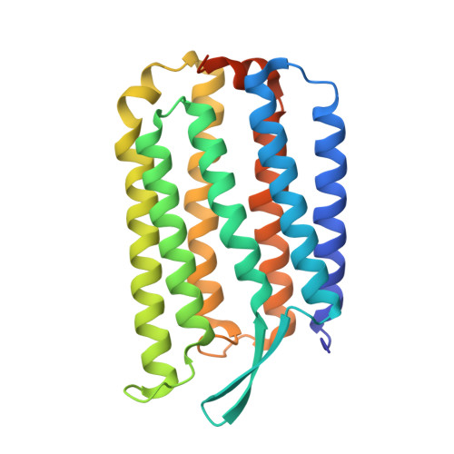
    Bacteriorhodopsins are a large family of seven-helical transmembrane proteins that function as light-driven proton pumps. Here, we present the crystal structure of a new member of the family, Haloarcula marismortui bacteriorhodopsin I (HmBRI) D94N mutant, at the resolution of 2.5 Å. While the HmBRI retinal-binding pocket and proton donor site are similar to those of other archaeal proton pumps, its proton release region is extended and contains additional water molecules. The protein's fold is reinforced by three novel inter-helical hydrogen bonds, two of which result from double substitutions relative to Halobacterium salinarum bacteriorhodopsin and other similar proteins. Despite the expression in Escherichia coli and consequent absence of native lipids, the protein assembles as a trimer in crystals. The unique extended loop between the helices D and E of HmBRI makes contacts with the adjacent protomer and appears to stabilize the interface. Many lipidic hydrophobic tail groups are discernible in the membrane region, and their positions are similar to those of archaeal isoprenoid lipids in the crystals of other proton pumps, isolated from native or native-like sources. All these features might explain the HmBRI properties and establish the protein as a novel model for the microbial rhodopsin proton pumping studies.


  • Organizational Affiliation
    • Institute of Complex Systems (ICS-6) Structural Biochemistry, Research Centre Jülich GmbH, Jülich, Germany; Laboratory for advanced studies of membrane proteins, Moscow institute of physics and technology, Dolgoprudniy, Russia.

Macromolecule Content 

  • Total Structure Weight: 33.88 kDa 
  • Atom Count: 2,044 
  • Modeled Residue Count: 236 
  • Deposited Residue Count: 258 
  • Unique protein chains: 1

Macromolecules

Find similar proteins by:|  3D Structure
Entity ID: 1
MoleculeChains  Sequence LengthOrganismDetailsImage
Bacteriorhodopsin258Haloarcula marismortui ATCC 43049Mutation(s): 1 
Gene Names: boprrnAC3161
Membrane Entity: Yes 
UniProt
Find proteins for Q5UXY6 (Haloarcula marismortui (strain ATCC 43049 / DSM 3752 / JCM 8966 / VKM B-1809))
Explore Q5UXY6 
Go to UniProtKB:  Q5UXY6
Entity Groups
Sequence Clusters30% Identity50% Identity70% Identity90% Identity95% Identity100% Identity
UniProt GroupQ5UXY6
Sequence Annotations
Expand
Reference Sequence

Small Molecules

Ligands 3 Unique
IDChains Name / Formula / InChI Key2D Diagram3D Interactions
RET

Query on RET


Download:Ideal Coordinates CCD File
B [auth A]RETINAL
C20 H28 O
NCYCYZXNIZJOKI-OVSJKPMPSA-N
LFA

Query on LFA


Download:Ideal Coordinates CCD File
C [auth A]
D [auth A]
E [auth A]
F [auth A]
G [auth A]
C [auth A],
D [auth A],
E [auth A],
F [auth A],
G [auth A],
H [auth A],
I [auth A],
J [auth A],
K [auth A],
L [auth A],
M [auth A],
N [auth A],
O [auth A],
P [auth A],
Q [auth A],
R [auth A],
U [auth A],
V [auth A],
W [auth A]
EICOSANE
C20 H42
CBFCDTFDPHXCNY-UHFFFAOYSA-N
SO4

Query on SO4


Download:Ideal Coordinates CCD File
S [auth A],
T [auth A]
SULFATE ION
O4 S
QAOWNCQODCNURD-UHFFFAOYSA-L

Experimental Data & Validation

Experimental Data

  • Method: X-RAY DIFFRACTION
  • Resolution: 2.50 Å
  • R-Value Free:  0.237 (Depositor), 0.239 (DCC) 
  • R-Value Work:  0.192 (Depositor), 0.196 (DCC) 
  • R-Value Observed: 0.194 (Depositor) 
Space Group: P 3 2 1
Unit Cell:
Length ( Å )Angle ( ˚ )
a = 102.854α = 90
b = 102.854β = 90
c = 60.003γ = 120
Software Package:
Software NamePurpose
MOLREPphasing
PHENIXrefinement
MOSFLMdata reduction
SCALAdata scaling

Structure Validation

View Full Validation Report


Ligand Structure Quality Assessment 


Entry History 

Deposition Data

Revision History  (Full details and data files)

  • Version 1.0: 2014-12-17
    Type: Initial release
  • Version 1.1: 2015-06-17
    Changes: Non-polymer description
  • Version 1.2: 2023-09-20
    Changes: Data collection, Database references, Derived calculations, Refinement description
  • Version 1.3: 2024-10-16
    Changes: Structure summary